International Journal of Anatomical Sciences 2010, 1:11-13
Research Paper
A Study of Microstructure of the Annular Ligament
Anand A.
Department of Anatomy, VMKV Medical College, Salem.
Key Words: annular ligament, cartilage, collagen fibers, cadaveric study
Abstract: In the elderly pain around the lateral side of the elbow is often diagnosed as lateral epicondylitis and the wear and tear of the common extensor origin is often implicated. However surgical ablation of the common extensor origin, physiotherapy and repeated intra articular injection have failed to alleviate the misery. Of late erosion of the inner surface annular ligament is considered to be a significant cause. Haematoxylin and eosin stained sections of the cadaveric annular ligament were studied to investigate the nature of the inner surface of the annular ligament. The inner surface of the annular ligament is lined by white fibrocartilage. The nature of the cartilage does not seem to be mentioned in standard references. The findings are discussed in detail.
The Annular ligament of the superior radio ulnar joint also known as the orbicular ligament in clinical practice is a very unique structure which forms four fifths of the articulating surface for the head of the radius. It plays a significant role in the stability of the superior radio ulnar joint (Tubbs et al., 2006). It is a resilient and a funnel shaped structure which has an outer and inner surface (Kanagasuntheram et al.,1996). The functional importance of annular ligament has not been comprehensively defined in orthopaedic literature (Bozkurt et al., 2005). The outer surface blends with the radial collateral ligament of the elbow and provides origin to the supinator muscle. The inner surface is lined by a thin coating of cartilage (Williams et al., 1986) and towards the lower aspect it is lined by synovial membrane. No other ligament has this uniqueness which truly makes it a pliable articulating surface. Standard textbooks of Anatomy do not mention the nature of this cartilage which lines this inner surface. The aim of the study was to ascertain the nature of the cartilage microscopically as well as look for any variations and macroscopic changes which could provide a clue for indeterminate causes for pain around the elbow for an orthopaedician (Faro and Wolf,2007).
Correspondance to: Anand A, Department of Anatomy, VMKV Medical College, Seeragapadi, Salem 636308, Tamilnadu, India.
Email: [email protected]
Materials and Methods
This study was carried out in 72 upper extremities belonging to both sexes after completion of dissection by the undergraduate students. Macroscopic dissections were performed on the specimens. The annular ligament was detached at both upper and lower ends from its osseous attachments. The inner surface of the annular ligament was examined for pitting, erosions and degeneration similar to bony changes. Tissue samples for microscopic studies were obtained with a predilection towards the upper part of the annular ligament. Tissues obtained were processed and paraffin blocks were prepared. Sections were taken from the aforesaid blocks and stained with haematoxylin and eosin stains. Slides were examined microscopically and lysed samples were excluded from the purpose of study.
Observations
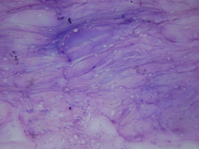
The study showed that all the ligaments examined were thick with a mean of 1.5 mm. The internal surface of the ligaments exhibited erosive changes indicative of wear and tear. In 32 specimens there was pitting similar to that of in bony articulating surfaces (Fig. 1). There was no evidence of degeneration. Histological examination of stained tissues exhibited features of dense fibrous tissue arranged in bundles with fibroblasts interspersed and small chondrocytes arranged in groups (Fig.2) amongst the bundles suggestive of white fibrocartilage. A total number of 54 stained tissues were examined microscopically and all showed evidence of white fibrocartilage (Fig. 2).
Fig. 1 Shows pitting on the inner surface of Annular ligament.
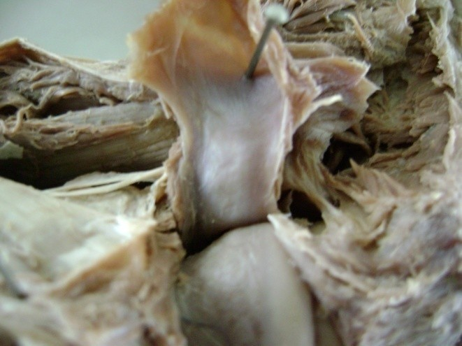
Fig. 2 Shows bundles of collagen fibers and fibroblasts.
The superior radio ulnar joint has the same fundamental structure as other synovial joints as it has a capsule, which is lined by synovial membrane and reinforced externally by accessory ligaments. The capsule of the elbow joint is in continuation with the capsule of the superior radio ulnar joint and the synovial membrane also being a continuity. The articular cartilages of synovial joints are formed of a special variety of hyaline cartilage which reflects their preformation as cartilaginous models in embryonic life (Barnett et al., 1961). The articular surfaces of bones which ossify in mesenchyme are covered with white fibrocartilage (Williams et al., 1986). In a synovial articulation the opposing articulating surfaces are usually hyaline cartilage in nature. When the opposing articulating surfaces possess different cartilaginous surfaces it tends to ossify as age advances. The sacro iliac joint was said to have such opposing articulating surfaces but it now conclusively proven that both auricular surfaces of the sacrum and the innominate bone are made up of hyaline cartilage (Standring and Mahadevan, 2008). Standard textbooks mention the articulating surface of the superior radio ulnar joint as osseo ligamentous ring but in view of evidence of white fibrocartilage it is safe to conclude and name it as a osseo cartilaginous ring of the superior radio ulnar joint.
Conclusion
White fibrocartilage is resistant to degeneration but is subject to vagrancy of time where it could undergo wear and tear but as age advances it tends to show a fibrous union restricting mobility which could contribute as a cause for chronic pain around the lateral side of the elbow. Hence practicing physicians have to bear in mind the cartilaginous nature of the annular ligament as a contributory factor for unresolved pain on the lateral side of the elbow.
Acknowledgements
I wish to express my sincere thanks and gratitude to my teacher and guide Prof. Melani Rajendran, HOD of department of Anatomy, SRMC & RI for permitting me to utilize the resources for dissections and Prof. Deepti Shastri, HOD of department of Anatomy, VMKVMC for permitting me to utilize the resources for dissections, histological studies and for constant guidance and encouragement.
References
Barnett CH, Davies DV, MacConaill MA (1961) Synovial joints, their structure and mechanics. London: Longmans. 63-64.
Bozkurt M, Halil Ibrahim A, Apaydin N (2005) The annular ligament, An anatomical study. Am J of Sports Medicine 33: 114-115.
Faro F, Wolf JM (2007) Lateral epicondylitis: review and current concepts. Am J of Hand Surgery 32:1271-1279.
Kanagasuntheram R, Sivananda Singam P, Krishnamurti A (1996) Textbook of Anatomy. Madras: Orient Longman Ltd. 88-89.
Standring S, Mahadevan V (2008) In: Gray’s Anatomy, The Anatomical Basis of Clinical Practice. 40th Edition, London: Churchill Livingstone Publishers. 1366.
Tubbs RS, Shoja MM, Khaki AA (2006) The morphology and function of the quadrate ligament. Via Medica, 65: 225-227.
Williams PL, Warwick R (1986) In:Gray’s Anatomy,36th Edition, London: Churchill Livingstone Publishers. 462.
Williams PL, Warwick R (1986) In: Gray’s Anatomy,36th Edition, London: Churchill Livingstone Publishers. 425-427.