International Journal of Anatomical Sciences 2010,1:39-41.
Case Report
Rare Origin of the Right Internal Thoracic Artery from Thyrocervical Trunk
Babu BP.
Department of Anatomy, Kasturba Medical College, Manipal 576 104, Karnataka, India.
Key Words: internal thoracic artery, thyrocervical trunk
Abstract: Rare origin of the right internal thoracic artery from thyrocervical trunk was observed in one of the 100 cadavers in the Department of Anatomy, Kasturba Medical College, Manipal, Karnataka during the period 2001-2009. An attempt has been made to compare this finding with earlier reports and also to highlight the clinical significance of the variation.
An uncommon origin of the right internal thoracic artery (ITA) from the thyrocervical trunk was observed in dissection of the root of neck. The Internal thoracic artery normally arises as a branch from the inferior aspect of first part of the subclavian artery. It courses downwards in the ventral thoracic wall and divides into two terminal branches in the sixth intercostal space, the musculophrenic and superior epigastric arteries (Gabella, 1995). It also supplies sternum, ventral thoracic wall and diaphragm through sternal, anterior intercostals and two terminal branches. Daseler and Anson (1959), in describing its origin state that it arises ventrally from the first part of the subclavian artery, inferior to thyrocervical trunk, just medial to scalenus anterior muscle. In their study on 769 specimens they found the internal thoracic artery arising as a direct branch from the third part of the subclavian artery in six (0.78%) of the specimens investigated.
Krechowiecki et al., (1973), in a study of 100 cadavers (200 arteries) on the variations of the course of the internal thoracic artery, found the artery originating lateral to scalenus anterior muscle in one case (0.5%).
Nizanowski et al., (1982), in their study on the ramifications of the subclavian artery, state that the vertebral and internal thoracic arteries showed the least deviation in their origin from the subclavian artery and abnormal or absent internal thoracic arteries in 11.4% of cases studied.
The Internal Thoracic Artery (ITA) has been widely used as a conduit in Coronary Artery Bypass Grafting (CABG) and revascularization of myocardium by surgical anastamosis of coronary artery and internal thoracic artery (Kuniyoshi et al.,
2002). The anatomy of internal thoracic artery facilitates its mobilization during surgery and its increasing use in coronary artery bypass grafting highlights the need to understand its variations. Internal thoracic artery arises from third part of subclavian artery in 0.83% of cases (Vorster et al.,1998) or from the thyrocervical trunk in more than 10% of cases (Lischka et al.,1982). In a rare case, as reported by Omar et al. (2000), ITA originated from third part of subclavian artery bilaterally, and yet in another case it gave origin to suprascapular artery (Yucel et al., 1999).
Case Report
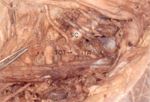
During routine dissection of head and neck in a male cadaver, the right ITA was found to arise from the thyrocervical trunk a branch from the first part of the subclavian artery (Fig. 1). The artery descended down and divided at the level of the right sixth costal cartilage into musculophrenic and superior epigastric arteries. The trunk of ITA provided the sternal branches, anterior intercostal arteries. No other abnormality could be detected.
Fig. 1 Photograph of the root of neck showing origin of right internal thoracic artery from Thyrocervical trunk.
(ITA-Internal thoracic artery; SC-Subclavian artery; TCT – Thyro-cervical trunk)
Discussion
The present case reports a rare origin of internal thoracic artery from the thyrocervical trunk on the right side in one of the 100 cadavers dissected (1%). Daseler and Anson (1959) found it in six (four Rt. & two Lt.) of 769 arteries (0.78%). Krenchowiecki et al., (1973) found it in one of 200 arteries (0.5%).
The anomalies found in the branches of the subclavian artery could be explained by considering the embryologic development of this region. The two factors influencing the development of these branches are the ability of the blood to follow the longitudinal channels offering least resistance and the tension on the vessels resulting from the caudal shifting of the heart and aorta.
Congdon (1922), in describing the development of the subclavian arteries from the seventh paired dorsal segmented arteries, found that tension on the distal portion of the right aortic arch causes that portion of the aortic arch and the right fourth dorsal segmental artery to form the proximal part of the right subclavian artery. With failure of the part distal to the seventh dorsal segmental branch to obliterate, degeneration of the proximal part occurs, resulting in abnormal origins of the right subclavian artery from the aortic arch or descending aorta.
The vertical part of the internal thoracic artery develops from ventral anastomoses between ventral divisions of thoracic intersegmental arteries. Due to the caudal shifting of the aorta, the proximal parts of these segmental arteries are exposed to longitudinal tension and bending with a resulting retarded blood flow. This may result in abnormal connections between the longitudinal channels (vertebral and internal thoracic arteries) and subclavian artery or aortic arch.
Conclusion
The internal thoracic artery is being used as alternative for revascularization of the myocardium in patients with coronary artery disease. Root of the neck is one of the sites commonly used in patients for percutaneous subclavian vein catheterization to determine central venous pressure (CVP) or to administer drugs and solutions in emergencies and for introducing a pacemaker.It is therefore important to be aware of this rare variation in the origin and course of the internal thoracic artery.
Acknowledgments
Author thanks Professor and Head of the Department of Anatomy for giving permission to publish the paper.
References
Congdon ED (1922) Transformation of the aortic- arch system during the development of human embryo. Contrib Embryol Carnegie Inst, 68: 47-110.
Gabella G (1995) Cardiovascular system. In: Williams PL, Bannister LH, Berry MM, Collins P, Dyson M, Dussek JE, Fergusson MWJ (Eds.). Grays’ Anatomy. 8th edition. London: Churchill Livingstone. 1534-1535.
Daseler EH, Anson BJ (1959) Surgical anatomy of the subclavian artery and its branches. Surg. Gynecol Obstet 108: 149-174.
Krechowiecki A, Daniel B, Wiechowski S (1973) Variation of the internal thoracic artery. Folia Morphol 32: 173-184.
Kuniyoshi Y, Koja K, Miyagi K, Uezu T, Yamashiro S, Arakaki K, Mabuni K, Senaha S (2002) Surgical treatment of aortic arch aneurysm combined with coronary artery stenosis. Ann Thorac Cardiovasc Surg, 8: 369-373.
Lischka MF, Krammer EB, Rath T, Riedl M, Ellbock E (1982) The human thyrocervical trunk: configuration and variability reinvestigated. Anat Embryol 163: 389-401.
Nizanowski C, Noczynski L, Suder E (1982) Variability of the origin of ramifications of the subclavian artery in humans (Studies on the Polish population). Folia Morphol 41: 281-294.
Omar Y, Lachman N, Satyapal KS (2001) Bilateral origin of the internal thoracic artery from the third part of the subclavian artery: a case report. Surg Radiol Anat, 23: 127-129.
Vorster W, Plooy Pt D, Meiring JH (1998) Abnormal origin of internal thoracic and vertebral arteries. Clin Anat, 11: 33-37.
Yucel AH, Kizilkanat E, Ozdemir CO (1999) The variations of the subclavian artery and its branches. OkajimasFolia Anat Jpn, 76: 255-261.

