International Journal of Anatomical Sciences 2010, 1: 42-44.
Case Report
An Unusual Flexor Digitorum Brevis Muscle and its Clinical Significance – A Case Report
Jayakumari S.
Department of Anatomy, Melmaruvathur Adhiparasakthi Institute of Medical Sciences and
Research, Melmaruvathur 603 319, Tamilnadu, India.
Key Words: flexor digitorum brevis, tendon
Abstract: An unusual flexor digitorun brevis muscle (FDB) was detected in the left foot of a 50 year old male cadaver. The muscle originated from the medial process of the calcanean tuberosity and was found to give off three tendons. The three tendons of FDB could be traced to second, third and fourth toes respectively. However, the tendon for the fifth toe was absent. Knowledge of such anatomical variants may be important clinically in view of reconstruction of heel pad by FDB musculocutanous flap transfer and interpretationof advanced diagnostic imaging modalities.
The flexor digitorum brevis (FDB) muscle is located between the abductor hallucis and the abductor digiti minimi and together they constitute the first layer of muscles on the sole. These muscles are closest to the skin of the sole superficially, surrounded by the planter aponeurosis and adjoined by the flexor digitorun longus (FDL) and the quadratus plantae. The muscle (FDB) takes origin from medial process of the calcaneus and plantar aponeurosis and inserted into the middle phalanges of second to fifth toes (Standring 2005).
The variations of the FDB were observed in 63% of cases, involving mostly the fifth toe and less fequently the fourth toe (Nathen and Gloobe 1973). Absence of fifth digit tendon was reported in 63.7% of cases (Chaney et al., 1996). Nathan and Gloobe (1973) noticed the absence of the fourth or most lateral slip of the muscle to the fifth digit in 23% of cases and in some of these cases, the absent tendon was replaced by a tendinous slip either from the flexor digitorum accessorius muscle or from the flexor digitorum longus muscle. The present study reports a variation in the composition of tendons of FDB and attempts to discuss its clinical implications. Awareness of anatomical variants of FDB is clinically relevent for performing reconstruction of heel pad and while using advanced diagnostic imaging modalities such as computed topography and magnetic resonance imaging.
Case Report
A variant of flexor digitorum brevis muscle was encountered in the left foot of a 50 year old male cadaver during the course of undergraduate medical training programme. The muscle originated from the medial process of the calcanean tuberosity, the central part of plantar aponeurosis and the intermuscular septa between it and adjacent muscles. It displayed a variation in the composition of its tendon. The muscle belly was found to give off three tendons near the mid segment of the foot. These three tendons were traceable to second, third and fourth toes respectively. However, the tendon for the fifth toe was absent (Fig.1).
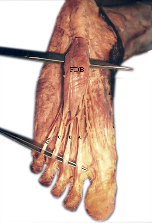
Fig. 1 Absence of flexor digitorum tendon for the fifth toe.
(FDB -Flexor Digitorum brevis muscle; FDL- Flexor Digitorum longus; A,B,C- tendon of flexor digitorum brevis muscle)
The muscle belly was 10.5 cm long and its maximum width was 2.5 cm. The tendons proceeding to second, third and fourth toes measured 9.4 cm, 6.8 cm and 5.8 cm respectively in length. The medial most tendon was relatively thin as compared to other two tendons. These tendons of FDB were found to be inserted as usual into the middle phalanx of the respective toes. The innervation of the muscle was derived from the medial planter nerve. There were no interconnecting bands between the individual tendons of FDB or with the tendon of flexor digitorum longus. No other morphological anomaly was noticed in the foot. Dissection of the right foot revealed normal anatomy of various soft tissue structures.
Discussion:
The knowledge of the normal and abnormal anatomy is essential in treating congenital abnormalities, traumatic and other pathological conditions of the foot and ankle (Chaney et al., 1996). FDB is a highly specialized muscle which helps to control the changes in the posture of the foot (Grogono and Jowsey 1965). It carries out flexion of second to fifth toes, a function maintainted by the FDL when the former is expanded (Hartrampt et al., 1980).
Standring (2005) reported the absence of fifth toe tendon, which may be replaced by small muscular slip from the long flexor tendon of from flexor accessorious. In the present case, absence of FDB tendon to little toe may possibly render tendon of FDL of that toe vulnerable to injury and displacement, since it is relatively unsupported. The neurovascular bundle along lateral side of the sole is liable to injury during surgical intervation in this region since it is deprived of the security provide by the fifth toe tendon FDB.
Three types of insertion of the FDB were recognized: absence of tendon, unsplit tendon, or tendon fused to the long flexor. The comparative assessment of size of the tendons of FDB revealed that the medial two tendons are usually larger than the lateral two (Sarrofian 1983). The present study revealed that the lengths of three tendons of FDB exhibited a decline from medial to lateral side and although the medial most tendon was the longest but it was found to be thinner than the remaining two tendons.
The toes are held extended at the metatarsophalangeal and distal inter- phalangeal joints and flexed at the proximal interphalangeal joints four toes. This probably results from the contraction of the terminal phalanges towards the sole and passively buckling the rest of the toes into the above position (Standring 2005). In the present case, presumably the flexion of little toe will be compromised because it will solely be carried out by FDL.
Reconstruction of plantar defects, especially the planter heel, presents a difficult problem as one of the main functions of this area is weight-bearing and reconstruction needs more anatomical consideration. The skin-grafted FDB flap provides an effective, feasible and reliable alternative reconstruction for extensive avulsed planter defects (Lin et al., 1991).
An understanding and awareness of the possible and predictable anatomical variants can prevent confusion during surgery and diagnostic testing. Familiarity with these variants is essential to prevent errors in the interpretation of advanced imaging techniques.
Reference:
Chaney DM,Lee MS,Khan MA,Krueger Wa,Mandracchia,VJ and Yoho RM (1996) Study of ten anatomical variants of the foot and ankle. J Am Podiatric Med Assoc, 86;11,532-537.
Grogono BJS, Jowsey J (1965) Flexor accessoreus longus.an unusal Muscle Anomaly. J Bone Joint Surg. 47: 118-119.
Hartrampf CR, Scheflan M and Bostwick J (1980) The flexor digitorum brevis muscle Island pedicle flap: A new dimension in heel reconstruction. Plast Reconstr Surgy, 66: 264-270.
Lin SD, Chou CK, Yang CC, Lai CS (1991) Reconstruction of planter heel defect using reinnervated, skin-grafted flexor digitorum brevis flap. Br J Plast Surg, .44: 109-112.
Sarrofian S (1983) In anatomy of foot and ankle. 2nd Edition, Philadelphia: J B Lippin Cott. 221-223.
Standring S (2005) In Gray’s Anatomy, 39th Edition, New York:Churcill Livingston. 1498-1499.