International Journal of Anatomical Sciences 2013, 4(2):26-29
Case Report
A Rare Case of Bilateral Small Kidneys in an Elderly Female – A Case Report
Arun Kumar S Bilodi, Jyoti D Kadam, Arjun Bahaddur, Gangadhar MR.
Department of Anatomy, Velammal Medical College Hospital and Research Institute Anupannadi post, Madurai – 625 009, Tamil Nadu, India.
Department of Radiology, Velammal Medical College Hospital and Reseach Institute Anupannadi post ,Madurai – 625 009, Tamil Nadu, India.
Department of Anthropology, University of Mysore, Manasa Gangothri, Mysore, Karnataka, India.
Key words: Renal aplasia, renal agenesis, bilateral small kidneys, solitary kidneys
Abstract: The objective present study is to report a rare case of bilateral small kidneys in elderly female age 75 years. This study was done in Bangalore district Bangalore. This was studied was during the month of April 2013. An elderly female aged 75 years old underwent general examination as outpatient at St .John’s Medical College Hospital for her extreme weakness heaviness in the chest and breathlessness since one month. A detailed history revealed that she is non diabetic, non hypertensive, and not suffering from ischemic heart disease. There was a definite family history of congenital renal anomalies .Her second daughter aged 50 years has absence of right kidney since birth. Her elder son had Horse shoe shaped kidneys associated with diabetes and hypertension, Thorough examination was done here and investigated She underwent all investigation like complete haemogram, 2D ECHO and ultrasound of abdomen which revealed Bilateral small kidneys with grade 1 parenchymal changes and left renal exophytic cyst. Renal agenesis and renal aplasia are known to cause anomalies of kidneys like congenital solitary kidneys which are more prone for renal failure. Familial history of anomalies and associated anomalies are known to cause higher incidence of renal agenesis.than renal aplasia . There is difficulty in differential diagnosis of renal agenesis and renal aplasia.It is said Renal agenesis which is diagnosed clinically is more due to renal aplasia. This study on bilateral small kidneys is rare may due to renal aplasia which is diagnosed clinically by ultrasonographically, which is very rare entity; hence studied and reported.
Severe abnormalities of kidneys are due to renal agenesis and renal dysplasia that causes diseases which requires either dialysis or renal transplantation in the first year of life. Classical example is multi-cystic diseases of kidney where nephrons fail to develop and ureteric bud fails to branch. If there is failure of interaction occurs between metanephric mesoderm and ureteric bud occurs, then renal agenesis occurs. Renal agenesis also occurs when there is mutation of genes that regulates expression of signaling of GDNF1. There may be absence of one or both kidneys. It is called as Agenesis. There may be underdevelopment of one or both kidneys known as Hypoplasia of kidneys. There may be over development of kidney known as Hyperplasia of kidney. Sometimes there may be presence of adrenal tissue. In some cases, there may be distention of pelvis occurs due to urine resulting in urinary passages obstruction. This condition is known as hydronephrosis (Singh and Pal, 2007).
Case Report
An elderly female aged 75 years came to outpatient department of cardiology at St. John’s Medical College Hospital for her extreme weakness heaviness in the chest and breathlessness since one month. A detailed history of personal history, drug history, past history, family history, history similar episodes in the past were taken. She was non diabetic, non hypertensive, and not suffering from ischemic heart disease. But there was definite family history of renal Anomalies. Her second daughter aged 50 years has absence of right kidney since birth, her elder son had Horse shoe shaped kidneys associated with diabetes and hypertension, Later thorough general and local examination were done here and investigated So she underwent relevant investigations like complete haemogram, 2D ECHO, X-Ray of the chest and ultrasound of abdomen were also done which revealed small bilateral kidneys with grade 1 parenchymal changes and left renal exophytic cyst.
Ultrasound of abdomen was done here at St John’s Medical College Hospital which is as follows:
i. Liver span is 10.6cm with no focal lesion
ii. Gall bladder is distended with CBD -3.5 cm, no caliculi
iii. Spleen is normal in size measuring 6.7 cm with no focal lesions
iv. Pancreas is normal with no duct dilatation.
v. Peripancreatic region is normal.
vi. No lymphadenopathy in the para aortic space.
vii. Kidney: The measurement of kidneys are as follows:
| DESCRIPTIONS | RIGHT KIDNEY | LEFT KIDNEY |
| Bipolar length | 8.1cm | 7.2cm |
| Parenchymal thickness | 0.8cm | 0.9cm |
Bilateral kidneys are smaller in size with grade 1 renal parencymal changes. Left kidney shows exophytic simple cyst measuring 2.3 cm x 2.2 cm in the upper pole of left kidney (left renal exophytic cyst). Urinary bladder–minimally distended. No free fluid in the abdomen.
Fig. 1 Ultrasonogram pictures showing small kidneys.

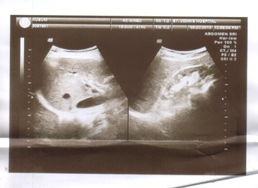
(Ultrasound photographs shows
o Both kidneys are small in size
o Subtle Obscuration and cortico medullary differentiation.
o Upper pole cortical cyst and size is 2.3cm and 2.2cmseen in left kidney.
o No evidence of any calculus nor pelvicalceal dilatations
o Right kidney measures 8.1cm in length,. Left kidney measures 7.2 cm)
Discussion:
During life, kidneys are reddish brown organs measuring 10cm in length, 5 cm in width, and 2.5 cm in thickness and moves with respiration of 2-3 cm in the vertical direction during the movement of diaphragm.
Primary screening was done in 52 babies for suspicion of small kidneys and one baby for multicystic kidney. But there was no renal agenesis. After one month, again second ultra sound was done .Three babies showed small kidneys with further decrease in size .They had no function .So they were diagnosed Renal Aplasia The incidence of Renal Aplasia is 1 in 1300. Again ultrasound was done which showed further regression in all three babies .By the end of one year kidney were hardly seen. These regressions may be due to Renal Aplasia (Masahiro et al. 2002). It is the genetic mutation or environmental factors that cause derangement in expression of genes (Woolf and Winnyard, 1998). In the autopsies studies, the most important cause of congenital solitary kidneys is Renal Agenesis (Fortune, 1927). A mother of two male children aged 45 years has absence of right kidney since birth There was no other anomaly in her body. Ultrasound of the abdomen showed on visualization of right kidney –Empty right renal fossa (Bilodi and Gangadhar, 2012). Her mother now has bilateral small kidneys since birth. DMSA Renoscintigraphy was done in nine children. There were 8 children with small kidneys & one with Multicystic dysplastic kidney. There were 4 kidneys which were non functioning, one showed bilateral hypoplasic kidney, four patient showed unilateral hypoplastic kidney. All five with hypoplastic kidneys were boys. No other congenital anomalies were present in these children. While passing urine, cystourethro-graphy showed association of vesico urethral reflex in all hypoplastic kidneys (Masahiro et al., 2002). There may be genetical relation between Renal genesis, renal aplasia and multicystic dysplastic kidney (MCDK) (Taxy, 1985). These anomalies are found in the members of same families (Bankier et al, 1985). Aplastic kidneys do have shape of reniform. They are small at birth but not rudimentary. These findings suggest that aplastic kidneys do grow almost to normal size. They have abnormal architecture. MCDK is not an end stage organ but said highly active in terms of genetic expression (Woolf and Winyard, 2000).
Retrospective evaluation was done to differentiate between scintigraphy patterns of congenital reflux nephropathy from that of acquired scarring in children with primary vesicoureteral reflux. They retro-specttively evaluated the frequency and pattern of renal scintigraphy abnormalities in 41 patients with prenatally detected primary vesicoureteral reflux. Dimercapto-succinic acid scintigraphy had been performed on 4-6 and 1-4 months. There was prenatal defect of vesico urethral reflex. Three types of renal damage were diagnosed. There was decreased uptake of renal radionuclide in 20-40% followed by, focal defects in uptake and shrunken kidney with relative uptake less than 20%. Scintigraphy revealed renal damage in 12 prenatally detected cases of vesicoureteral reflux, including decreased uptake in 58% and shrunken kidney in 42%, and in 111 cases of reflux detected at urinary tract infection (Polito et al., 2000).
In the present study, an elderly female aged 75 years has bilateral small kidney as per ultrasound reported She is not diabetic nor hypertensive. She has no any clinical symptoms involvement renal system. All her parameters were normal; but she had mild degree of incompetence of valves in her heart No history of ischemic heart disease but there was strong family history of anomalies of renal system. She is mother of 5 children; out of them two has congenital anomalies of renal system. Her elder son had horse shoe shaped kidney, while her elder daughter has an absence of right kidney. So this condition of bilateral small kidneys may be due to shrinking that might have taken place during the antenatal period. These types of kidneys are known as aplastic kidneys which may be due to mutation of genes or may be due to environmental factors in causing the derangement of genes. So, she has these bilateral small kidneys.
Conclusion
This study reports about bilateral shrunken kidney. It may be renal aplasia which is having an incidence of 1 in 1300. Being a rare anomaly, it has been studied and reported.
Reference:
Sadler TW (2010) Langman’s Medical Embryology: South Asian Edition. 11th Edition. Lippincort: Williams & Wilkins. p240.
Singh IB, Pal GP (2007) Human Enbryology. 8th edition. Delhi: Macmillan India Ltd. p244.
Moore KL, Dalley AF, Agur AMR (2010) Abdomen – Chapter 2 In: Clinically Oriented Anatomy: Lippincort: William & Willkins. Walters Kluwer Health. p292.
Masahiro H, Hirokazu T, Yuusei O, Kenkou K, Yashinori I, Mitsufumi M (2002) Renal aplasia is the predominant cause of congenital solitary kidneys. Kidney Intern, 61:1840-1844.
Woolf A, Winnyard P (1998) Advances in the cell biology & genetics of the Human Body Malformations. J Am Soc Nephrol, 9: 1114-1125.
Fortune C (1927) The pathological and clinical significance of congenital one sided kidney defect with presentation of three new cases of agensia & one of aplasia. Ann Intern Med, I:377-399.
Bilodi AKS, Gangadhar MR (2012) A Case of unilateral renal agenesis in a female. A case report. Int J Anat Sci, 3: 04-07.
Taxy J (1985) Renal Dysplasia: A review. Pathol Ann, 20:139-159.
Bankier A, Decampo M, Newell R (1985) A pedigree study of perinatally lethal renal disease J Med Genet, 22: 104-111.
Woolf AS, Winyard PJ (2000) Gene expression and cell turn over in human renal dysplasia. Histol Histopathol, 15: 159-166.
Polito C, Monnaa LA, Rambald PF, Nappi B, Mansi L, DiToro R (2000) High Incidence of a generally small kidney and primary vesicouretal reflux. J Urol, 164: 479-482.