International Journal of Anatomical Sciences 2011,2(1):7-10
A Study on the Variations of Median Nerve formation in South Indian Population
Sarala Devi KV, Udhaya K, Sudha Seshayan
Department of Anatomy, Vinayaka Mission’s Kirupanandha Variyar Medical College, Salem 636 308, Tamilnadu, India
Registrar, Tamilnadu Dr.M.G.R. Medical University, Chennai, India.
Key Words: Median nerve, lateral cord, musculocutaneous nerve
Abstract: The median nerve is one of the commonest nerve showing variations from the level of its formation to its termination. This is an important region where anesthetist, plastic surgeon and oncologist land up with problems more frequently due to variations and the mismanagement will result if they are not aware of these variations. The median nerve was studied regarding its formation in 60 upper limb specimens by conventional dissection method. Two specimens showed variations in our study. In one specimen, low level formation of median nerve with innervation of arm muscles was found. In another specimen the median nerve was formed at a higher level by the fusion of medial root with lateral cord of brachial plexus. These findings will provide anatomical knowledge for the reliable clinical correlation and surgeries.
Median nerve is formed by the union of lateral root from lateral cord (C5, C6 and C7) and medial root from medial cord (C8 and T1) of brachial plexus lateral or antero-lateral to the 3rd part of axillary artery (Turner, 1864; Williams et al.,1999; McMinn, 1990). Variations in the formation of median nerve are common, more frequent and have been observed by different authors in India (Satyanarayana et al., 2010; Krishnamurthy et al., 2007). Such study in south Indian population is comparatively sparse. Knowledge of these variations is essential for performing nerve repair, nerve transplant and reconstructive surgeries. Hence this study would be of great help to the plastic surgeons and orthopedic surgeons. Hence we undertook
Correspondence to: K.V. Sarala Devi, Department of Anatomy, Vinayaka Mission’s Kirupanandha Variyar Medical College, Salem, Tamilnadu. India Email: sarala[email protected]
Accepted :15-April-2011 this study to find out the variations in formation of median nerve in South Indian population.
Materials and Methods
Sixty upper limb specimens from30 embalmed human cadavers of South Indian population irrespective of sex were used for the present study. They were dissected by conventional dissection method (Romanes, 1986) and examined for anatomical variations in median nerve formation.
Observations
The median nerve showed two variations in two specimens (out of 60 specimens). No associated muscular and vascular anomaly was noticed.
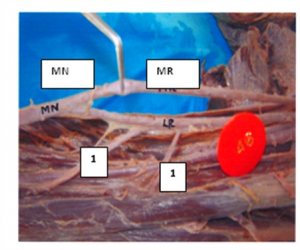
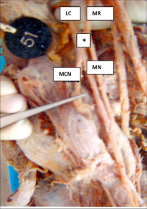
Fig 1: Low level formation of median nerve
 |
 |
LC – lateral cord; MR – medial root; MN–median nerve; MCN – musculocutaneous nerve; * – site of formation of median nerve (accidentally it was splitted when dissecting)
One specimen (fig. 1) showed the formation of median nerve at the junction of middle and lower 1/3rd of arm. The medial root of median nerve was longer than usual (13.6 cm). The lateral cord of brachial plexus after giving the lateral pectoral branch, continued down in the arm. The medial root crossed in front of brachial artery from medial to lateral side and then united with the lateral cord to form the trunk of median nerve. The musculocutaneous nerve was absent. The lateral cord supplied branches to coraco- brachialis, biceps brachii, and brachialis. This anomaly was unilateral and the incidence was 1.66%.
One specimen (Fig. 2) was noticed with median nerve formation at a higher level than usual. After the lateral pectoral nerve was given off, lateral cord of brachial plexus was joined by medial root to form median nerve. About 0.7cm distal to the formation of median nerve, mus- culocutaneous nerve was given off. It was presenting unilaterally and the incidence was 1.66%
Discussion
Anatomical variations of median nerve are frequent and there are many studies reported. Chauhan and Roy (2002) reported the formation of median nerve by two lateral roots and one medial root. Satyanarayana et al. (2008) and Uzun and Seelig (2001) noted that the median nerve was formed by four roots, three lateral and one medial root. In other study, it was observed that musculocutaneous nerve was absent and so the median nerve supplied muscular branches to coracobrachialis, brachialis and biceps brachii (Guha and Palit, 2005). Satyanarayana et al. (2010) also reported that formation of median nerve by three roots, two from lateral cord and one from medial cord. In another study, the C7 fibers were traced from the median nerve to the musculocutaneous nerve (Walsh, 1877). In our study the median nerve was contributed by the lateral cord and root from medial cord but the musculocutaneous nerve was given off from median nerve; the evidence of this anomaly was found to be less in previous literature. The surgeon has to keep these variations in mind especially during radical axillary dissection and shoulder arthroscopy wherever sparing of the nerve is mandatory
The incidence of distal formation of median nerve was more common (8.5%, Uysal et al., 2003; 12%, Matejcik 2003;2.1%, Mohammed and Badawoud, 2003) than that of high level formation. Due to these neural variations, sometimes the arm muscles are innervated by median nerve and in most of such cases, musculo- cutaneous nerve was found to be absent (Prasada Rao and Chaudhary, 2000; Song et al., 2003, Satheesha nayak 2007, Beheiry 2004). In contrast to our study, Melani Rajendran and Nivedha, (2004) noted that the median nerve in addition to its formation in the middle of arm, gave off muscular branches to arm muscles and musculocutaneous nerve. The finding of our present study coincides with that of Satheesha nayak (2007). The knowledge of these variations of median nerve should be borne in mind during evaluation of unexplained nerve palsy after trauma, surgical intervention of particular area, neurotization of neural injuries and in reconstructive surgeries
In humans, the muscles of the upper limb develop from the mesenchyme of the paraxial mesoderm during the 5th week of embryonic life (Larsen, 1977). The neurons penetrate into the mesen- chyme in different direction (Brown et al.,1991; Williams et al., 1995). Immediatelyenter the limb buds and establish an intimate contact with the differentiating mesoderm condensations. This early contact between the nerve and muscle cells is a prerequisite for their complete functional differentiation (Saddler, 2006). These variations of the nerves are the result of alterations in signaling between mesenchymal cells and neuronal growth cones during development which once formed could persist postnatally (Sannes et al., 2000). Probably, the neurons of the nerve in our study would have also taken such anomaly.
Conclusion
These neural variations should be thought of while managing recurrent neuropathies with unusual clinical symptoms and signs. This is an important region where the anesthetists, plastic surgeons, oncologists and radiologists land up with problems frequently due to variations and mismanagement will result if they are not aware of these anatomical variations.
References
Beheiry EE (2004). Anatomical variations of the median nerve distribution and communication in the arm. Folia Morphol (Warsz) 63: 13-18.
Brown MC, Hopkins WG, Keynes, RJ (1991) Essentials of neural development. In: Axon guidance and target recognition. Cambridge University Press. Cambridge. pp 46 – 66.
Chauhan R, Roy TS (2002) Communication between the median and musculocutaneous nerve – a case report. JASI 52: 72–75.
Guha R, Palit S (2005) A rare variation of anomalous median nerve with absent musculocutaneous nerve and high up division of brachial artery. J Interacad 9: 398–403.
Krishnamurthy A, Nayak SR, Venkatraya Prabhu L, Hedge RP, Surendran S, Kumar M, Pai MM (2007) The branching pattern and communications of musculocutaneous nerve. J Hand Surg Eur 32: 560-562.
Matejcik V (2003) Anatomic deviations in the origin of the median nerve. Ro zhl chir82:526-528.
McMinn RMH (1990) Upperlimb. In: Last’s Anatomy. 8th Edition. Churchill Livingstone, Edinburgh. pp 82.
Melani Rajendran S, Nivedha R (2004) An anomalous median nerve. Anatomical Adjuncts. 4: 23-25.
Sarala Devi, et al.., Variation of Median Nerve Formation in South Indian Population
Badawoud MHM (2003) A study on the anatomical variations of median nerve formation. Bahrain Medical Bulletin 25: 1-5.
Prasada Rao PV, Chaudhary SC (2000) Communication of the musculocutaneous nerve with median nerve. East Afr Med J 77:498-503.
Romanes GJ (1986) Cunningham’s Manual of Practical Anatomy. Vol I 15th Edition, Oxford University Press, London. pp 67-74.
Saddler TW (2006) Muscular system. In: Langman’s Medical Embryology. 10th Edition. Lippincott Williams & Wilkins, Philadelphia. pp 146–147.
Sannes HD, Reh TA, Harris WA (2000) Development of the nervous system In: Axon growth and guidance. Academic Press, New York. pp 189-197.
Satyanarayana N, Guha R (2008) Formation of median nerve by four roots. J College of Medical Sci-Nepal, 5: 105–107.
Satyanarayana N, Reddy CK, Sunitha P, Jayasri N, Nitin V, Praveen G, Guha R, Datta AK, Shaik MM (2010) Formation of median nerve by three roots – a case report. J College of Medical Sci-Nepal 6: 47-50.
Satheesha Nayak (2007) Absence of musculo- cutaneous nerve associated with clinically important variations in the formation, course and distribution of the median nerve – a case report. Neuroanatomy 6: 40–50.
Song WC, Jung HS, Kim HT, Shin C, Lee BY, Koh KS (2003) A variation of the musculo- cutaneous nerve absent. Yonsei Med J, 44:1110–1113.
Turner WM (1864) On some variations in the arrangements of the nerves of the human body. Natural History Review 4: 612–617.
Uysal II, Sekar M, Kasabulut AK, Buyukmumlu M, Ziylan T (2003) Brachial plexus variations in human fetuses. Neurosurg, 53: 676-684.
Uzun A, Seelig LL Jr (2001) A variation in the formation of the median nerve; communicating branch between the musculocutaneous and median nerves in man. Folia Morphol (Warsz)80: 99–101.
Walsh JF (1877) The Anatomy of the brachial plexus, Am J Med Sci, 74: 387-397.
Williams PL, Bannister LH, Berry MM, Collins P, Dyson M, Dussek JE, Ferguson D (1995) Nervous system In: Gray’s Anatomy. 38th Edition. Churchill Livingston, Edinburgh. pp 1266–1274.
Williams PL, Bannister LH, Berry MM, Collins P, Dyson M, Dussek JE, Ferguson D (1995) Embryology and development In: Gray’s Anatomy. 38th Edition. Churchill Livingstone, Edinburgh. pp 231–232.
