International Journal of Anatomical Sciences 2010, 1: 17-20
Research Paper
Greater Splanchnic Nerve
Swayam Jothi S, Hemanth K, Ravi Kumar U, Rajeswara Rao N.
Shri Sathya Sai Medical College and Research Institute, Kanchipuram, Tamilnadu, India
Key Words: greater splanchnic nerve, sympathectomy.
Abstract: Sympathectomy is done for relief of pain concerned with abdominal organs such as pancreatic pain. According to Naidoo (2001) establishing a predictable pattern of splanchnic neural anatomy may be of surgical relevance. Therefore an attempt has been made to study the level of origin as well as the pattern of formation of the greater splanchnic nerve in 25 adult cadavers with a view to obtain more information about them to help overcome the failure rate in sympathectomy procedures. The highest level of origin was seen from T4 and the lowest ganglia contributing to the formation of the greater splanchnic nerve was T11.
Sympathetic preganglionic efferent fibres emerge through the thoracic and upper lumbar spinal nerves as white rami communicans and reach the sympathetic chain. These fibres emerge from the medial side of lower thoracic sympathetic chain to form the splanchnic nerves. The greater splanchnic nerve usually is formed by the myelinated preganglionic efferents emerging via 5 to 9 or 10th thoracic ganglia. They end in the coeliac ganglia.
Sympathectomy is done for relief of pain concerned with abdominal organs such as pancreatic pain. Srickland (1995) tried performance of local anesthetic and placebo splanchnic nerve blocks via indwelling catheter to predict benefit from thoracic splanchnectomy in a patient with intractable pancreatic pain. According to Naidoo (2001) establishing a predictable pattern of splanchnic neural anatomy may be of surgical relevance. Therefore an attempt has been made to study the level of origin as well as the pattern of formation of the greater splancnic nerve in 25 adult cadavers with a view to obtain more information about them to help overcome the failure rate in sympathectomy procedures.
Correspondance to: Swayam Jothi S, Shri Sathya Sai Medical College & Research Institute, Kanchipuram, Tamilnadu, India. Email:[email protected]
Material & Methods
During dissection of thorax after the removal of the lungs the sympathetic chain was cleaned and the ganglia were defined. The origin of the greater splanchnic nerve from different ganglia and its formation were noted.
Observation
The number of ganglia from which the greater splanchnic nerve was arising varied from 2 ganglia to 6 ganglia and in one the greater splanchnic nerve was absent. (Table 1).
Table 1
| No. ofspecimens | Total no. of gangliainvolved in formation |
| 1 specimen | 6 ganglia |
| 7 specimens | 5 ganglia |
| 17 specimens | 4 ganglia |
| 22 specimens | 3 ganglia |
| 2 specimens | 2 ganglia |
| 1 specimen | Absent |
The level of origin of the greater splanchnic nerve varied from the T4 – T11 ganglia. The highest level of origin was from T4 ganglion (Fig. 1). The lowest level of origin was fromT11 (Fig. 2).
Fig. 1 Highest level of origin of greater splanchnic nerve from T4 thoracic ganglion (GSN – Greater splanchnic nerve )
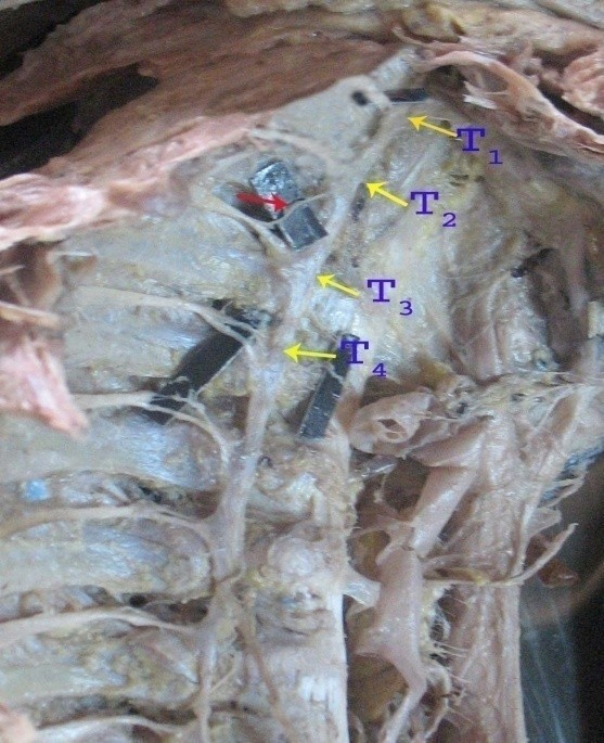
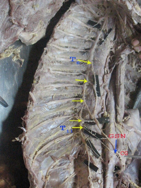
Fig. 2 Lowest level of origin of greater splanchnic nerve from T11 sympathetic ganglion. (GSN – Greater splanchnic nerve)
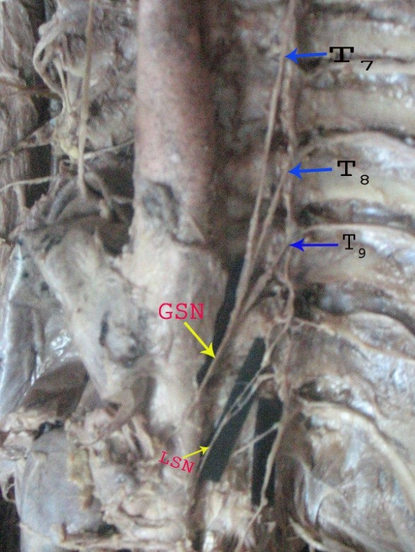
The most common origin was from three ganglia (Fig. 3) as seen in 22 specimens (Table 1).
Fig. 3 Pattern of origin of greater splanchnic nerve from 3 ganglia (T7,8,9). (GSN – Greater splanchnic nerve; LSN – Lesser splanchnic nerve)
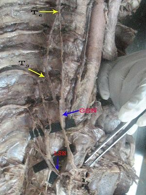
Maximum number of a specific pattern was observed in the case of 4 ganglia (Fig. 4) where the origin was from 6,7,8,9 thoracic ganglia.
Fig. 4 Pattern of origin of greater splanchnic nerve from 4 thoracic ganglia (T6, 7, 8, & 9) (GSN – Greater splanchnic nerve; LSN –Lesser splanchnic nerve)
This was present in 10 specimens (Table 5). The pattern of origin within the specified number of ganglia also varied. (Table 2).
Table 2
|
The details of the patterns are given in Table 3, 4, 5, 6 & 7. Two patterns were observed when the greater splanchnic nerve was arising from 2 ganglia (Table 3). Nine patterns were seen when it was arising from 3 ganglia (Table 4). Five patterns were seen when it was arising from 4 ganglia (Table 5) Two patterns were seen when it was arising from 5 ganglia (Table 6). Only one pattern was seen when it was arising from 6 ganglia (Table 7).
|
Table 3
Table 4
| Patterns | No of specimens |
| 5,6,7 |
01 |
| 5,7,9 |
01 |
| 6,7,8 |
03 |
| 6,7,9 |
03 |
| 6,8,9 |
04 |
| 6,8,10 |
03 |
| 7,8,9 |
05 |
| 7,9,10 |
01 |
| 8,9,10 |
01 |
Table 5
| Patterns | No of specimens |
| 4,6,7,8 |
01 |
| 5,6,7,8, |
03 |
| 5,7,8,9 |
01 |
| 6,7,8,9 |
10 |
| 6,8,9,10 |
02 |
Table 6
| Patterns | No of specimens |
| 5,6,7,8,9 |
04 |
| 6,7,8,9,10 |
03 |
|
Table 7
Discussion:
The greater splanchnic nerve showed great variability both in the level of its origin and in the pattern of its formation. Reed (1951) found 58 patterns of the origin of the greater splanchnic nerve among 100 cadavers. In the present study 19 patterns of origin were seen in 50 specimens. Edward and Baker (1940) found the greater splanchnic nerve arising from 3 ganglia in only 23% of the bodies as compared to the findings of the present study which shows 44% (22 specimens). Moreover, the splanchnic nerves were rarely bilaterally symmetrical according to him. In the present observation, it was bilaterally symmetrical in three cadavers.
The observation that the greater splanchnic nerve arising from T7,T8,T9 ganglia in 5 specimens is similar to the observations of Edward and Baker (1940) who have stated that the most frequent origin in their series was from the seventh, eighth, and ninth ganglia.
Reed (1951) in 100 bodies (both sides) found the highest contribution to the greater splanchnic nerve came from the fourth ganglia in 4 cases; from fifth ganglion in 64 cases; from the 6th ganglion in 85 cases; from the seventh ganglion in 41 cases and from the eighth in 6 cases. In the present study the highest origin of the greater splanchnic nerve from the 4th ganglion was found in one specimen and the lowest from the 11th ganglion in one specimen.
Among many patterns of arrangement which Reed (1951) observed, the most common pattern was seen in 13 specimens (6.5%) in which the greater splanchnic nerve was formed by the rami from the sixth, eighth, ninth thoracic sympathetic ganglia. In the present study, the large number of the greater splanchnic nerves with a common pattern was seen in 10 specimens (20%) and the rami were coming from 6th 7th 8th and 9th ganglia.
Swayamjothi et al., Greater splanchnic nerve
From these observations it is obvious that in thoracolumbar sympathectomy the sympathetic chain has to be removed upto the highest point of origin of greater splanchnic nerve to overcome failure rate in sympathectomy.
References:
Edwards LF, Baker RC (1940) Variation in the formation of the splanchnic nerves in the man. Anat Rec, 77: 335.
Naidoo N, Pratab P, Pather N, Moodley J, Singh B, Satyapal KS (2001) Thoracic splanchnic nerve; implication for the splanchnic denervation. J Anat, 199: 585-590.
Reed AF (1951) The origins of the splanchnic nerves. Anat Rec, 109:341.
Srickland T (1995) Performance of local anaesthetic and placebo splanchnic blocks via indwelling catheter to predict benefit from thoracoscopic splanchnectomy in a patient with intractable pancreatic pain. Anesthesiology, 84: 980-983.