International Journal of Anatomical Sciences 2012, 3(1): 01-03
Research Article
Abnormal Origin of Vertebral Artery
Anitha, V.
Department of Anatomy, Kanyakumari Government Medical College, Asaripallam, Kanyakumari 629 201, Tamil Nadu, India.
Key words : vertebral artery, arch of aorta, artery
Abstract: Abnormal origin of vertebral artery (left) from arch of aorta was noticed during dissection of fifty specimens and the same is presented for its rarity and clinical significance.
The vertebral artery takes origin from the first part of subclavian artery and its branches supply the spinal cord, medulla oblongata and cerebellum.
It has become important to know the exact origin and course of the vertebral artery as well as the percentage of abnormalities of the variations from the point of view of surgery, angiography and in all non invasive procedures and for subsequent stabilization procedures of the cervical spine. Thorough knowledge of the anatomy of vertebral artery is mandatory to avoid potentially catastrophic injury to the vertebral artery.
Materials and Methods
The extra cranial course of vertebral artery was dissected in 25 formalin fixed cadavers (both sides) in Institute of Anatomy, Madras Medical College. It included 17 male and 6 female cadavers and 2 fetal cadavers. Careful dissection was carried out to find the details of variations on both sides and photographs were taken. Observation
The vertebral artery (left) originated directly from arch of aorta in one adult specimen (2%). The right side vertebral artery originated from the first part of sub- clavian artery in the same specimen (Fig. 1).
In the remaining specimens, the vertebral artery was found to originate from the first part of subclavian artery in the scaleno vertebral triangle.
Discussion
Gray (1901), Piersol (1930), Grant (1951), Romanes (1964) have reported that the vertebral artery arises from the first part of subclavian artery.
Shivkumar et al., (2010) has found an anomalous branching pattern of the aortic arch while dissecting a 55 year old male cadaver in which the left vertebral artery originated from aortic arch proximal to the origin of left subclavian artery. Jeyanthi et al., (2010) has reported an anomalous origin of left vertebral artery directly from aortic arch in between left common carotid and left subclavian arteries
Fig. 1 Photograph showing abnormal origin of vertebral artery
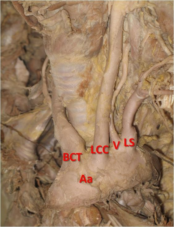 |
Aa-Aortic arch, BCT -brachiocephalic trunk, LCC-left common carotid, V-vertebral artery, LS- left subclavian Beeta Patasi et al., (2009) found an abnormal origin of left vertebral artery from arch of aorta between left common carotid and left subclavian arteries, during dissection of a 77 year old male cadaver.
|
|
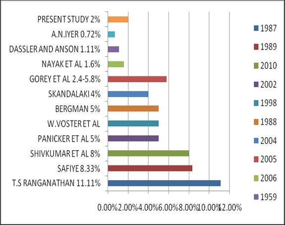
Fig. 2 Comparative study of origin of vertebral artery from arch of aorta.
Soubhagya and Nayak et al., (2006) reported in 1.6% of the 62 cadavers dissected that the left vertebral artery had its origin from arch of aorta.
Gorey et al., (2005) has stated that the most frequent variant is the left vertebral artery arising directly from arch of aorta between left common carotid and left subclavian arteries which accounts to about 2.4-5.8 percent.
Skandalaki (2004) has reported that in 4 percent of cases the vertebral artery originated from arch of aorta.
Panicker et al., (2002), Voster et al., (1998), have reported that the vertebral artery originated from arch of aorta in 5 percent of cases
Cavdar and Arisan (1989) has quoted that in 8.33 percent of cases, the left vertebral artery had its direct origin from arch of aorta.
Ranganathan and Jothi, (1987) has stated that the vertebral artery originated from arch of aorta in 11.11 percent of cases.
Bergman and Affifi (1988) have illustrated that the usual branches (80 percent) of arch of aorta were brachiocephalic trunk, left common carotid and left subclavian artery. If vertebral was present, more frequently it was left than the right vertebral. The variation of the four vessels has a frequency of about 5 percent.
Dassler and Anson (1959) have said that the left vertebral artery originated from the arch of aorta in 1.11 percent of the two bodies dissected.
Iyer (1917-1926) has recorded 6 out of 828 bodies (0.72%) in which the left vertebral artery originated from the arch of aorta.
In the present study, the origin of vertebral artery (left) from the arch of aorta was found in one specimen (2%) of the total fifty specimens dissected.
Conclusion
The vertebral artery originated from the arch of aorta in 2% of cases in present study.
Neurosurgical procedures like proximal brachial plexus repair, scalenotomy, direct isolation of proximal vertebral artery requires good working knowledge of the first segment of vertebral artery. In angiographic or anatomic post mortem examinations, abnormal vertebral artery origins are only incidental findings, because, for most of the cases they are clinically asymptomatic. Nonetheless, these abnormalities are of diagnostic importance either prior to vascular surgery in the neck region or in cases of intravascular diseases such as arteriovenous malformations or cerebral aneurysms.
Acknowledgement
Annual convocation of maortic arch distal to left subclavian artery. AJNR,26: 93-95.The author would like to thank Dr.Christilda Felicia, the then HOD of Anatomy, Madras Medical College, Chennai, for her support and valuable suggestions.
References
Berman RA, Afifi AK (1988) Anatomy Atlases: Illustrated Encyclopedia of Human Anatomic Variation: Opus II: Cardiovascular System: Arteries: Head, Neck, and Thorax, http://www.anatomyatlases.org/AnatomicVariant s/Cardiovascular/Text/Arteries/Aorta.shtml
Cavdar S, Arisan E (1989) Variations in extracranial origin of the human vertebral artery. Acta Anat (Basel), 135: 236-238.
Dasler EH, Anson BJ (1959) Surgical anatomy of subclavian artery and its branches. Surg Gnyecol Obst, 108: 149–174.
Gorey AR, Joshi R, Garg A, Merchant S, Yadav B, Maheswari P (2005) Aortic arch variation: a unique case with anomalous origin of both vertebral arteries as additional branches of the aorticarchdistaltoleftsubclavianartery.AJNR,26:93–95.
Grant JCB (1951) The Anatomy of the respiratory, blood vascular and lymphatic system. Oxford University Press, New York Gray H (1901) Anatomy – Descriptive and Surgical.
Philadelphia, Lea brothers
Iyer AA (1927) Some anomalies of origin of the vertebral artery. J Anat (originally J Anat Physiol), 62: 121–122.
Jeyanthi V, Prakash, Devi MN, Geethanjali BS, Rajini T (2010) Anomalous origin of the left vertebral artery from arch of aorta;Review of literature and a case report. Folia Morphol(warsz), 69: 258-260.
Panicker HK, Tarnekar A, Dhawane V, Gosh SK (2004) Anomalous origin of left vertebral artery:- Embryological basis and applied aspects-a case report. J Anat Soc India, 51: 234-235
Patasi B, Yeung A, Shannon Goodwin Aliroza Jalali SGA (2009) Anatomical variation of the origin of the left vertebral artery. Int J Anat Variations,2: 83-85.
Piersol GA (1930) Human Anatomy. 9th Edition. JB Lippincott Company, Philadelphia.
Ranganathan TS, Swayam Jothi S (1987) Variations of subclavian artery. 10 the Association of Anatomists at Stanley Medical College, Madras.
Romanes GJ (1964) Cunnighams’ Text Book of Anatomy. 10th edition. Oxford University Press.
Shivkumar GL, Pamidi N, Somayali SN, Nayak S, Vollala VR (2010) Anomalous branching pattern of the aortic arch and its clinical applications Singapore Med J, 51: e182-183.
Skandalakis JE (2004) The Embryologic and Anatomic Basis of Modern Surgery. Paschalidis Medical Publication Ltd.
Nayak SR, Pai MM, Prabhu LV, D’Costa S, Shetty P (2006) Anatomical organisation of aortic arch variations in India: Embryological basis and review. J Vascular Brasilerio, 5: 95-100.
Voster W, Du Plooy PJ, Meiring JH (1999) Abnormal internal thoracic and vertebral arteries. Clin Anat,1: 33-37