International Journal of Anatomical Sciences 2012, 3(1): 23-25
Research Article
Anatomy of Amorphic Branching Pattern of Brachial Artery and Its Clinical Complications – A Cadaveric Study
Priya G, Radhika Krishnan J, Sundarapandian S.
Department of Anatomy, SRM Medical College, Hospital and Research Centre, SRM Nagar, Potheri Village, Kattankulathur 603 203, Tamilnadu, India.
Key Words: brachioradial artery, vasa aberrantia, superficial ulnar artery, medial and lateral ulnar collateral arteries.
Abstract: The Information of an amorphic brachial artery branching patterns and incidence will be important for clinicians and surgeons when performing invasive procedures in arm and forearm. In this study ten cadaveric specimens were dissected, all were with an average age of 50-55 years out of which two cadavers showed high bifurcation of brachial artery seven was normal and one showed abnormal variations which is rare and not yet reported. The Branching pattern observed was the right brachial artery gave a branch just opposite to the origin of superior ulnar collateral and there was no inferior ulnar collateral so these arteries are named as medial and lateral ulnar collateral instead of later. Then just 1cm above the cubital fossa ulnar artery was observed which coursed superficially in the forearm as superficial ulnar artery. On the left side double brachial artery and which got joined by vasa aberrantia was found. This cadaveric survey may give awareness to anesthetists to prevent intra- arterial cannulation, drug administration and for orthopaedicians while operating the elbow injury. The abnormal anastomosis around elbow joint will be useful for microsurgeons.
This study was undertaken to determine the frequency of variations involved in the anatomy of branching pattern of brachial artery. A series of 10 embalmed cadavers were dissected and the revealed result was 4% incidence of high bifurcation,1% superficial ulnar artery and amorphic anastomoses around elbow joint, 1% Vasa aberrentia.
Materials and Methods
A total of 10 embalmed cadavers (20 upper limbs) were examined. The limbs had been partially dissected by SRM preclinical medical students and then further dissected by the authors.
Observation
High division of the brachial artery was observed in two cadavers. The brachial artery divides into its two terminal branches just below the origin of the Profunda brachii. The lateral branch was the radial artery, located in the space normally occupied by the brachial artery and the medial one was the ulnar artery.
An anomalous branching pattern of brachial artery was observed in one cadaver. The right brachial artery gave a branch just opposite to the origin of superior ulnar collateral which descends down on the coracobrachialis, brachialis muscle and behind brachial artery to the medial epicondyle to anastomose with anterior ulnar recurrent. There is no inferior ulnar collateral. So the branches are named as medial ulnar collateral (MUC) and lateral ulnar collateral (LUC) instead of superior and inferior (Fig2). In the cubital fossa 1cm proximal to the elbow ulnar artery was observed which coursed superficially in the forearm as superficial ulnar artery (SU) (Fig1) along with the basilic vein and with no branches.
Fig. 1 Cubital Fossa Showing Superficial Ulnar
Artery
|
|
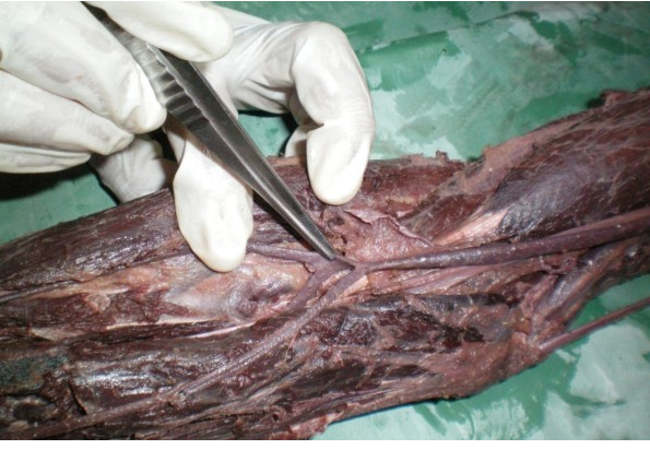
U – Superficial Ulnar Artery, IA – Interosseous
Artery, BA – Brachial Artery, RA – Radial Artery
Fig. 2 Arm Showing Anomalous Branches of Brachial Artery Named as Medial & Lateral Ulnar Collateral Artery
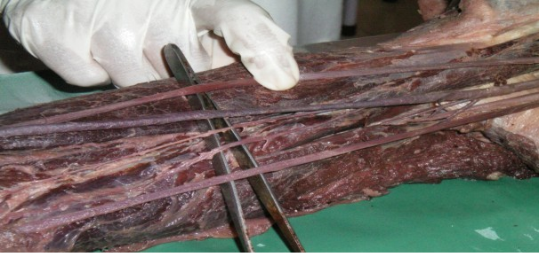
RA – Radial artery, LUC – Lateral Ulnar
Collateral, MUC – Medical Ulnar Collateral, MN
– Median Nerve, UN – Ulnar Nerve
Anastomosis around elbow joint
In front and back of the medial epicondyle of humerus, the lateral ulnar and medial ulnar collateral anastomosed with anterior and posterior divisions of ulnar recurrent artery which is a branch of common interosseous artery. On the lateral epicondyle the anastomosis was normal.
The left brachial artery after giving profunda brachii divided into two branches of same caliber one coursed superficial to the median nerve from medial to lateral and continued in the forearm as radial artery hence it is named as brachioradial artery. This artery is closely related to the cephalic vein and the other coursed as the normal brachial artery. In the cubital fossa brachioradial artery gave a communicating branch called Vasa aberrantia, which has joined brachial artery in an arch like pattern in front of the bicipital tendon.
Discussion
Anatomical variations in the arterial supply of the upper limb pose a hazard for inadvertent intra-arterial cannulation and drug administration (Chin and Singh, 2005). In two cadavers we observed high bifurcation of brachial artery, the lateral one resembled the normal course of brachial artery and medial is the ulnar but in one cadaver the lateral branch superficially crossed the median nerve and is closely related to cephalic vein. It could be easily mistaken for a vein during intravenous injection. Such misinterpretation could lead to intra-arterial injection. This can be minimized by careful palpation and Doppler flow meter (Devansh, 1996). There is a case report of gangrene of upper extremity followed by intra-arterial injection of drugs (Goldberg et al., 1984).
Superficial position of the ulnar artery makes it more vulnerable to trauma and this leads to hemorrhages and it also has a higher risk of getting damaged during forearm surgery (Devansh, 1996). The clinical manifestation depends on two factors; the location of the injury and presence of anatomical variations. In the case of the Biceps tendon rupture, the presence of Vasa aberrantia as in the present case may be injured which will lead to ischemic myositis, myoneurosis, impaired vascularity or claudication (Augustus et al.2008). If arterial injury is suspected arteriography should be obtained.
The anastomosis around the elbow joint is a rare finding here. The knowledge of such anastomosis recorded in this study will be useful for the micro-surgery and orthopaedic surgeons.
The dislocation of the elbow causes stretching and distortion of the anterior structure which involves disruption of circulation. The anatomy of arrangement of neurovascular structures in the elbow is important in both normal and variant. In this study there is 1% incidence of amorphic arrangement of vessels observed in the cubital fossa from lateral to medial:
1. Brachioradial artery (BRA)
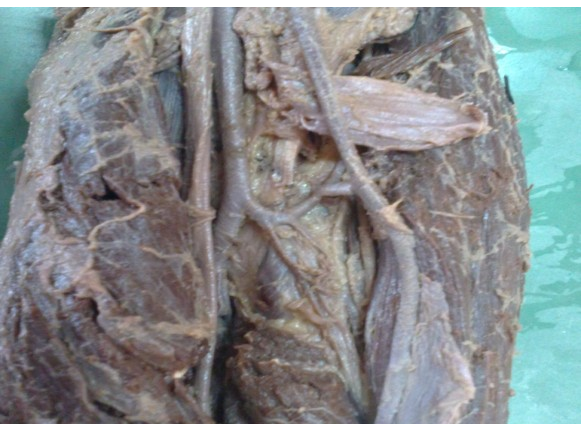
2. In the center Biceps tendon. Anterior to the tendon is Vasa aberrantia (VA) (Fig.3)
3. Medial to the tendon brachial artery(BA)
4. Medial nerve (MN)
Fig. 3 Cubital Fossa Showing Vasa Aberrantia pressures are obtained when the diagnosis is in doubt and arteriography is obtained (Augustus et al., 2008; Yamaguchi et al.,1997)
Medial elbow instability has become an increased recognized entity, particularly among throwing athletes. The fundamental knowledge of the medial elbow anatomy and vascular anatomy of the elbow is a must because there is a chance of rupture of the superficial ulnar artery. Injury to the artery may result in decreased hand circulation (Sean et al., 2008)
References
Augustus D, Mazzocca Md, Jeffery T, Spang MB, Robert A, Arciero MD (2008) Distal Biceps rupture. Orthoclin NAM, 39: 237-249.
Chin KJ, Singh K. (2005) The Superficial ulnar artery– potential hazard in patients with difficult venous access. Br J Anaestr, 94: 692-693.
Devansh (1996) Superficial ulnar artery flap. Plast reconstr surg, 97: 420-426.
Goldberg L, Bahar A, Yosipoirtch Z (1984) Cangrene of upper extremity following intra –arterial injection of drugs. A case report and review of the literature. Clin orthop relat Res, 188: 223-229.
Rodriguez–Niedenfuhr M, Vazquez T, Nearn L,
|
UA VA
SBA
Ferreira B, Parkin I, Sanudo Jr (2001) Variation of the arterial pattern in the upper limb revisited; a morphological and statistical study, with a review of the literature. J Anat, 199: 547-566.
Sean P, Grace MD, Larry D, Field MD (2008) Chronic Medial Elbow instability. Orthop clin AM, 39: 213-219.
Williams et al., (2005) Gray’s Anatomty 39th Edition pp393.
y, Yamaguchi K, Sweet FA, Bindra R et al. (1997) TheDBA – Brachial Artery, UA – Ulnar Artery, VA –
Vasa Aberrantia, RA – Radial Artery
Injury to the Vasa aberrantia causes compartment syndrome results in intra muscular bleeding and edema formation within the flexor compartment of the forearm. Pain with passive finger and wrist extension out of proportion to the injury raises clinical suspicion. Compartment extraosseous and intraosseous arterial anatomy of the adult elbow J Bone joint Surg AM, 79: 1653-1662.