International Journal of Anatomical Sciences 2012, 3(2): 36-38
Research Paper
Study on Struther’s Ligament with Supracondylar Spur / Process and its
Clinical Significance
Ganesan Murugaperumal†, Melani Rajendran S‡
Department of Anatomy, Tagore Medical College and Hospital, Rathnamangalam, Vandalore (PO), Chennai 600 048, Tamil Nadu, India.
Department of Anatomy, Sri Ramachandra Medical College and Research Institute, Porur, Chennai 600 116, Tamil Nadu, India.
Key words: Struthers’ ligament, Supracondylar process, pronator teres muscle, median nerve, brachial artery
Abstract: A study was carried out to find out the prevalence of Struthers’ ligament. Out of 30 upper limbs dissected, in one upper limb, a Struther’s ligament had been identified. Its attachments, relationships to a rudimentary supracondylar process, the pronator teres muscle, median nerve, brachial artery and the medial epicondyle of the humerus were studied.
Struthers’ ligament is an inconstant ligament (Varlam, 2005; Dellon, 1987; Hommel, 1989) that extends between the shaft of the humerus and the medial epicondyle of the humerus (De Jesus, 2003). Its clinical significance arises from the fact that the median nerve passes in the space between the ligament and the humerus, and in this space the nerve may be compressed leading to supracondylar process syndrome (Wertsch, 1982; Bilecenoglu, 2005). Median nerve compression at the wrist is a common problem. Entrapment at other locations below or just above the elbow is uncommon. Therefore the present study was carried out to find out the prevalence of Struthers’ ligament and its relation with median nerve, brachial artery and pronator teres muscle.
Materials and Method
The study was conducted in the Department of Anatomy, Sri Ramachandra Medical College and Research Institute, Sri Ramachandra University, Porur, Chennai. 30 upper limbs, belonging to 10 embalmed cadavers and 10 free upper limbs were used for the study. Out of 10 embalmed cadavers,6 were males and 4 were females. The dissection been done on the distal third of the arms of both sides. After the dissection of the ulnar nerve, the region was surveyed for the existence of musculotendinus and fibrous structures resembling an arch and related to any neurovascular structures. Data been recorded and the specimens with Struthers’ ligament were photographed.
Observations
Out of 30 upper limbs studied, only one free upper limb showed the presence of Struthers’ ligament and supracondylar spur/process in the lower one third of the arm (Fig. 1). The observation was found on a left free upper limb. The Struther’s ligament was found to be attached to the supra condylar spur of humerus. The Struther’s ligament was 3 cm and the supra condylar spur was 1 cm in length. From the ligament, the fleshy fibers of pronator teres arose besides its regular origin from the medial epicondyle (Fig. 2). The brachial artery and median nerve passed deep to the tendinous arch formed by Struther’s ligament and supracondylar spur in the lower one third of the arm (Fig. 1), instead of passing superficial to brachialis muscle.
Fig. 1 Supra condylar process and Struther’s ligament (lower and upper horizontal blue arrows)
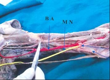
BA- Brachial Artery; MN- Median Nerve
Fig. 2 Fleshy fibers of pronator teres muscle
(horizontal blue arrow)
|
|
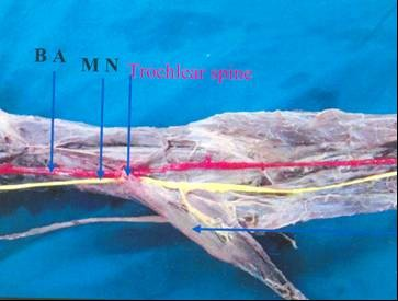
BA: Brachial artery, MN: median nerve Discussion
John Struthers, a Scottish anatomist, described several musculotendinous arches, the supracondylar spur/process, and the ligament between this process and the medial humeral epicondyle (the ligament of Struthers) at the distal third of the arm, and the relation of these structures to the median nerve and brachial artery (Struthers,1848). Knox in 1841 first reported its occurrence in man, as it was previously thought to be present only in animals (Newman, 1969). While the spur presents its relative frequency in the European Caucasians, the process and its homologous forms rarely occurred in early man and the anthropoids as well as in many of the lower apes.
Romanes, (1964) stated that the Struther’s ligament have to be borne in mind in performing venesection at the elbow. Sometimes the brachial artery accompanies the median nerve behind the supracondylar process of humerus from which a fibrous arch is most often thrown over the artery. This condition resembles the normal condition in some carnivores (Huber, 1930; Romanes, 1964; William et al., 1999). Occasionally the brachial artery is crossed in some part of its course by muscular or tendinous slips derived from coracobra- chialis, biceps, brachialis or pronator teres (Williams et al., 1999),
In the present study in one case (3.3%), Struther’s ligament and supratro- chlear spine were found in the lower one third of the arm from which fleshy fibers of pronator teres arose besides its origin. Median nerve and the brachial artery passed deep to the trochlear spine and struther’s ligament. This observation is in accordance with Hofer and Hofer (1910), who described a case in which brachial artery passed between the heads of pronator teres instead of dividing above it but they were unable to find a similar case in literature. Such cases can cause median nerve entrapment along with the brachial artery (Paval, 2008).
The supracondylar spur/process of the humerus is a rare skeletal anomaly, which is usually seen in an asymptomatic patient as a painless mass on an X-ray taken for some other purpose. The anatomic relationship with the neurovascular structures is well demonstrated by magnetic resonance imaging (MRI) (Lordan, 2005)
Melani and Ganesa – Struther’s ligament with supracondylar processThe process can fracture resulting in pain and tender mobile swelling over the medial aspect of the arm and consequent
It is essential to owe knowledge on supracondylar spur/process and Struther’s ligament since the presence of them may often associate with neurovascular distur- bances in the corresponding upper extremity.
Examination for the presence of a supracondylar spur should be considered during the approach to a patient of pain and sensory disturbance of the forearm and/or hand.
References
Bilecenoglu B, Uz A, Karalezli N (2005) Possible anatomic structures causing entrapment neuropathies of the median nerve: an anatomic study. Acta Orthop Belg, 71: 169–176.
De Jesus R, Dellon AL (2003) Historic origin of the “Arcade of Struthers”. J Hand Surg Am, 28: 528-531.
Dellon AL, Mackinnon SE (1987) Musculo- aponeurotic variations along the course of the median nerve in the proximal forearm. J Hand Surg [Br], 12: 359–363.
Hofer K, Hofer G (1910) Ueberden Verlauf der arteria brachialis mit dem nervus medianus zwischen den beiden kopfen des musculus pronator teres. Anatomical Anzeles, 36: 510.
Hommel U, Bellée H, Link M (1989) The validity of parameters in neonatal diagnosis and fetal monitoring of breech deliveries. 1. Neonatal status after breech delivery (in German). Zentralbl Gynakol, 111: 1293–1299.
Huber GC (1930) In:Piersol’s Human Anatomy. In: The vascular system, 9th Edn. Vol.I.J.B. Lippincott. Co. Philedelphia. pp767-791.
Lordan J, Rauh P, Spinner RJ (2005) The clinical anatomy of the supracondylar spur and the ligament of Struthers. Clin Anat, 18: 548-551.
Newman A (1969) The supracondylar process and its fracture. Am J Roentgenol, 105: 844-848.
Paval J (2008) A rare case of possible median nerve entrapment. Neuro Anatomy, 5: 35-38.
Arteries of the upperlimb. Walls, E.W.Edr.10
Edn. Oxiford University Press, New York: pp 885-893.
Struthers J (1848) On a peculiarity of the humerus and humeral artery. Mon J Med Sci, 28: 264-267.
Varlam H, St Antohe D, Chistol RO (2005) Supracondylar process and supratrochlear foramen of the humerus: a case report and a review of the literature (in French). Morphologie,89: 121–125.
Wertsch JJ, Melvin J (1982) Median nerve anatomy and entrapment syndromes: a review. Arch Phys Med Rehabil, 63: 623–627.
Williams PL, Bannister LH, Berry MM, Collins P, Dyson M, Dussek JE, Fergussion MWJ (1999) Gray’s Anatomy In: Cardio Vascular system. Gabella, G. Edr. 38th Edn. Churchill Livingstone. Edinburgh, London: 1542-44.