International Journal of Anatomical Sciences 2013, 4(2):20-22
Case Report
Branching Pattern of Arch of Aorta – A Case Report
Jacintha Antony, Rajeswara Rao N, Hemanth Kommuru, Swaym Jothi S.
Department of Anatomy, Sree Balaji Medical College & Hospital, Chromepet, Chennai – 600 044, Tamil Nadu, India.
Department of Anatomy, Shri Sathya Sai Medical College & Research Institute, Ammapettai, Nellikuppam – 603 108, Tamil Nadu, India.
Key words: aorta, aorta branching pattern
Abstract: An anomaly in the branching pattern of arch of aorta observed during routine dissection classes for medical graduates is reported here for its academic value and interest.
The arch of aorta begins behind the right border of the sternum at the level of the second costal cartilage and extends up to the left of body of fourth thoracic vertebra (McVay et al., 1984). The aortic arch gives rise to three classical branches the brachio cephalic trunk, left common carotid artery & left subclavian artery (Williams et al., 1989). Branches of the aorta vary in its origin but in some rare cases the position also varies. If it is high and is at the root of the neck, a most dangerous position in emergency tracheostomy. During the routine dissection of root of the neck and superior mediastinum the anomaly observed was reported.
Case Report
By careful dissection the arch of aorta and its branches are traced.
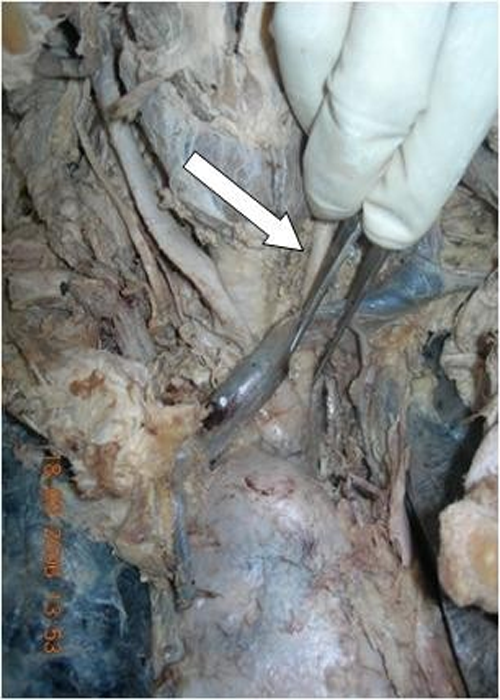
Left common carotid artery was arising in common with brachiocephalic trunk (Fig 1) and was crossing from the right to left side at the root of the neck (Fig 2). They were crossed by the left bra-chiocephalic vein in front. (Fig. 3). The crossing of the left common carotid artery took place 2.5 cm below the isthmus of the thyroid gland in front of the trachea about 1 cm above the suprasternal notch of the sternum (Fig 4).
Fig. 1 Shows Left common carotid artery arising in common with brachio-cephalic trunk from the arch of the aorta.
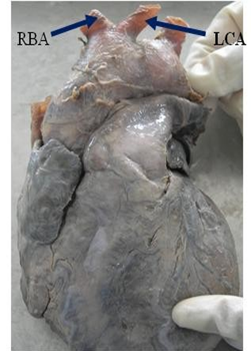
(RBA – Right brachiocephalic trunk LCA – Left common carotid artery)
Fig. 2 Shows left common carotid artery crossing from right side to the left side
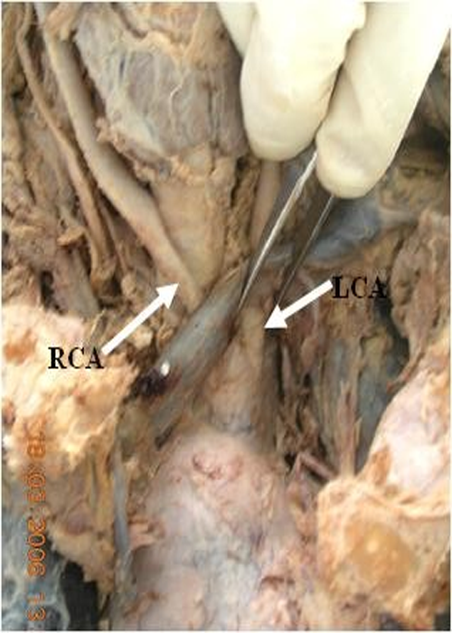
(RCA – Right common carotid artery LCA – Left common carotid artery)
Fig. 3 Common origin of brachiocephalic artery & left common carotid artery being crossed by left brachio-cephalic vein.
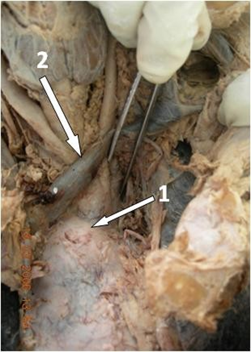
(1 – Left common carotid artery; 2 – Left brachiocephalic vein)
Fig. 4 Left common carotid artery crossing the trachea 2.5cms below the isthmus of the thyroid gland
Discussion:
As far as the branches of the aortic arch are concerned there are plenty of variations in their origins.
Anson et al. (1963) made an analysis of variations in the branches from thousand aortic arches and showed the following findings – The most common patterns 65% is formed by the separate origin of three branches springing from the vessel’s convex aspect, in 27%, the left common carotid artery originates from the brachiocephalic trunk. In 2.5% each of the four arteries originates independently from the arch of the aorta separately, the remaining showed a great variety of patterns, the commonest being 1.2% symmetrical right and left brachio cephalic trunks (Anson, 1963). According to Mc Donald and Anson (1940) the so called normal type of branching apparently occurs only in a little more than 50% of Negroes, but in almost 75% of Whites. The most common variation from this, said to occur approximately twice as often (in almost 40% of cases) in Negroes, is the origin of the innominate and the left common carotid artery together; the common stem for these may be extremely short, hardly more than a slight bulge in the aorta, or , rarely quite long (Williams et al., 1989).
Conclusion
Because of many changes involving in the transformation of the embryonic aortic arch system into the adult arterial pattern, it is understandable that variations may occur. This anomaly has to be remembered and care must be taken while doing emergency tracheostomy – a life saving maneuver. Additionally, a variant of origin and course of a great vessel arising from the aortic arch is of great clinical value, because of the ignorance on behalf of the surgeon of such a variation may cause serious surgical complications during procedures performed in the superior mediastinum and at the root of the neck.
Reference:
Anson BH (1963) The aortic arch and its branches In: Cardiology. Vol.1. New York: Mc Graw – Hill. p68.
Mc Donald JJ, Anson BJ (1940) Variation in the origin of arteries derived from the aortic arch, in American whites and negroes. Am J Phys Anthropol, 27: 91.
Mc Vay CB, Anson BJ (1984) Surgical anatomy. 6th Edition,Philadelphia: WB Saunders Co.426–433.
Williams PL, Warwick R, Dyson M, Bannister LH (1989) Gray’s Anatomy. 37th Edition. Edingburgh: Churchill Livinstone. 733–734.