International Journal of Anatomical Sciences 2013, 4(1):22-23
Case Report
INIENCEPHALY-A Case Report
Jacinta Antony, Swayam Jothi D, Sai Suchitra.
Department of Anatomy, Sree Balaji Medical College & Hospital, Chromepet, Chennai – 600 044, Tamilnadu, India.
Key words: Iniencephaly, inion, spina bifida, meningocoele
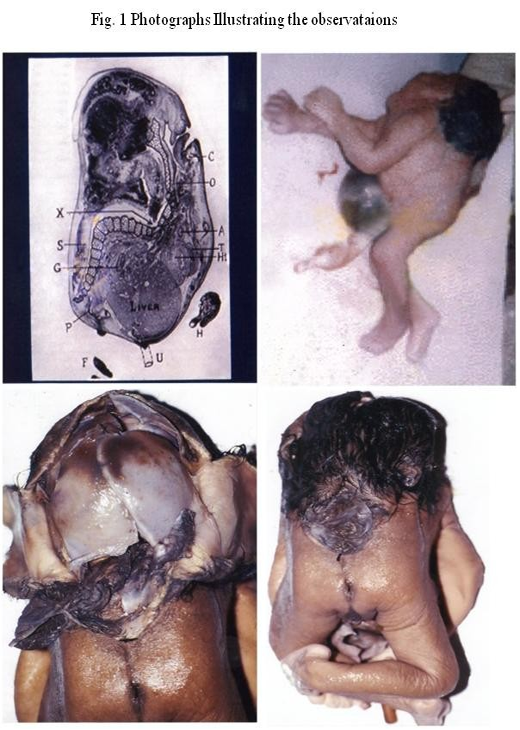
Abstract: A full term dead born foetus delivered by Caesarean Section from the Obstetrics and Gynaecology Department of the Government Kilpauk Medical College & Hospital was handed over to the Department of Anatomy for further study. The head of the foetus was retroflexed, a cystic swelling was present dorsally above the anal orifice and ventrally an omphalocoele.
Iniencephaly is a rare malformation which is incompatible for post-natal survival. It is more common in female sex than male and has been extensively described by Ballantyne (1904) and Gilmour (1941).
Case Report
The face of the foetus was well formed. The posterior hairline of the scalp merged with the skin of the thoracodorsal region thereby obliterating the nape of the neck. There was a cystic swelling above the anal orifice.
The scalp was opened by a coronal and a sagittal incision. The frontal and parietal bones were intact. The brain had autolysed. The squamous part of the occipital bone was absent. There was no foramen magnum. The base of the skull presented the cranial nerves in their respective foramina.
A mid sagittal section was made on the dorsal aspect encircling the cystic swelling. The laminae and spines in the lumbar vertebrae were absent-spina bifida. The vertebral canal contained the spinal cord and the swelling was a meningocoele. Omphalocoele was present ventrally.
Discussion:
The absence of the inion on the squamous part of the occipital bone which is absent is responsible for the title of this paper. This condition is normally associated with spina bifida or meningocoele.
Borderland and Morison in their text book on Foetal and Neonatal Pathology state that this condition cannot be distinguished from KLIPPER-FEIL SYNDROME. It has been reported as early as 1904.
Conclusion
Foetus with more than one anomaly is an interesting case for study. This requires study of each region in detail regarding its development and also the genetic reasons for such malformation. This condition is reported here because it had more anomalies than reported earlier. Past history and family history did not reveal any causative factor. The genetic abnormality leading to this condition could not be carried out.
References
Gilmour JR (1941) The essential identity of Klippel-Feil syndrome and iniencephaly. J Pathol, 53: 117-131.
Lewis HL (1897) Iniencephalus. Am J Obstet, 35: 11-53.
Morison JE (1904) Gross malformations. Neural tube and axial skeleton. P157.
Osborne DF (1948) Iniencephalus. Can Med Assoc J, 59: 474-475.
Willis RA (1958) The borderland of embryology and pathology. 2nd Edition. Chapter 4. 132-162.