International Journal of Anatomical Sciences 2013, 4(1):27-28
Case Report
High Origin of Radial Artery – A Case Report
Sai Sucheethra D, Sree Lekha D, Swayam Jothi Dorai Raj S, Rajeswara Rao N
Department of Anatomy, Guntur Medical College, Guntur – 522 004, Andra Pradesh, India.
Key words: radial artery, anomalous origin
Abstract: An unusually high origin of radial artery observed during routine dissections was reported here for its rarity.
Superficial arteries of the arm are relatively common. McCormack (1953) found some types in 30.77% of 364 bodies in which both limbs were investigated. They were commonly unilateral, so the incidence among limbs was much less (18.53% of 750 limbs).
During routine dissection two brachial arteries were observed in the right arm of a male cadaver. The arteries were traced proximally and distally.
Case Report
The following observations were made – The largest branch of brachial artery – the arteria profunda brachii was arising from the third part of the axillary artery in common with subscapular artery (Fig. 1).
Two brachial arteries were seen (Fig. 2) one (superficial brachial artery) was found in continuity with the radial artery at the level of cubital fossa (Fig. 3). The other had usual course and divided into its terminal branches namely radial and ulnar arteries at the level of the tuberosity of the radius.
|
|
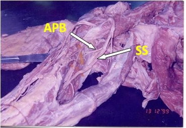
(APB – arteria profounda brachia, SS – Sub scapular artery)
Fig. 1 common origin of arteria profunda brachi and subscapular artery arising from 3rd part of axillary
|
|
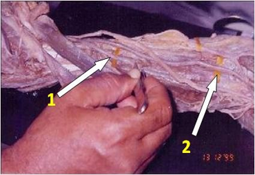
Fig. 2 Presence of two brachial arteries (1 & 2)
Discussion:
The primitive radial artery arises from the axial artery in the arm; where as the definitive radial artery arises from the axial artery at the level of the bend of the elbow. Subsequently the primitive radial artery joins with the definitive radial artery. At a later date the primary radial regresses completely but occasionally it may persist so as to account for the high origin of the radial artery from the brachial artery in the arm.
|
|
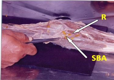
(SBA–superficial brachial artery communicating with the radial artery, R- Radial artery)
Fig. 3 Deeply placed brachial artery divided into ulnar & radial arteries. Superficial brachial artery was communicating with radial artery
Trotter and her colleagues (1934) found the subscapular, both circumflex scapular, the profounda brachii and the superior ulnar collateral all share a common stem, the axillary artery then bifurcating into radial and ulnar arteries.
Origin of a superficial brachial artery in the arm formed 5.57 percent of the total number of vascular variations they observed in these limbs. High origin of radial artery formed 77% of all variations of the arteries of the arm in 12.14% it arose from the brachial artery 2.66% of limb has an anastamotic connections with the brachial artery (McCormack and Co workers, 1953)
In the present case the brachial artery was doubled. The high origin of radial artery was due to the persistence of the primitive radial artery. This anomaly could be explained with an embryological background. Further knowledge about the variation will be of use while reducing fracture of the humerus, in reparative surgery and during cardiac catheterization.
Reference:
McCormack LJ, Cauldwell EW, Anson BJ (1953) Brachial and ante-branchial arterial patterns: A study of 750 extremities. Surg Gynec & Obst, 96:43.
Trotter M (1934) Septal apertures in the humerus of American whites and Negroes. Am J Phys Anthropol, 19:213.