International Journal of Anatomical Sciences 2014, 5(2): 46-48.
Case Report
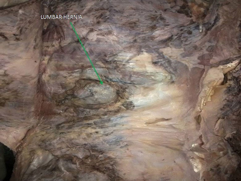
Lumbar hernia- A cadaveric Case Study
Kirubhanand C, Tamil Selvi P, Vijaya Prakash KM, Shradha Iddalgave,
Department of Anatomy, Karuna Medical College, Vilayodi, Palakkad- 678 103, Kerala, India.
Department of Anatomy, Sathyabama Dental College & Hospital, Sathyabama University, Chennai 600 119, Tamil Nadu, India.
Department of Anatomy, Dr. ALM PGIBMS, University of Madras, Taramani Campus, Chennai 600 113
Key words: lumbar hernia, petits triangle
Abstract: In this article we report a case of cadaveric finding of inferior lumbar hernia. Lumbar triangle hernia that occurs through lumbar triangles is very rare type of hernia. Only about 300 cases have been reported till date. Petit’s triangle hernia find further rarity and the case under reference is probably the first ever reported case of Petit’s triangle hernia in cadaveric finding. The relevant literature has been reviewed and the case report is discussed in brief.
Lumbar hernias are quite uncommon as compared to other ventral abdominal wall hernias, accounting for less than 1.5% of all abdominal hernias, with fewer than 300 cases reported over the past 300 years. About 25% of all lumbar hernias have a traumatic etiology (Bhasin and Khan, 2006). Lumbar hernia can occur individually or in association with certain syndromes or following trauma. This may be post-surgical or following blunt injuries associated with intra-abdominal injuries (Walgamage et al., 2015). Clinical diagnosis of this entity is difficult due to non-specific symptoms. The diagnosis is particularly elusive in obese individuals or in post-surgical patients. Though rare defects, lumbar hernias are prone to incarceration and strangulation
(Grauls et al., 2004).
The swelling may appear on coughing and disappear on compressing it. On clinical examination there may be single circular/oval swelling of 10 x 8 cms arising from the inferior lumbar triangle with an expansile impulse on pressurizing over lateral abdominal wall. It may be tender or non-tender and reducible on compression (Michael and Richardson, 2012).
Lumbar hernias are rare defects that involve the extrusion of retroperitoneal fat or viscera through a weakness in the posterolateral abdominal wall. Within this region there are two anatomically defined weaker triangles, the triangle of Petit and the triangle of Grynfelt-Lesshaf. The “triangle of Petit” or “the inferior lumbar triangle” is an upright triangle bound by the crista ilica, the musculus obliquus externus and the musculus latissimus dorsi (Singh and Kumar, 2014). The superior lumbar triangle is an inverted triangle bordered by the 12th rib, the musculus serratus posterior inferior, the musculus quadratus lumborum, the musculus erector spinae and the musculus obliquus internus. Lumbar hernia may be asymptomatic, associated with a sense of discomfort, or the cause of notable localized tenderness. – lumbar hernias are rare, the differential diagnosis must be made with a lipoma, a soft tissue tumor, a hematoma, an abscess, an atheromatous cyst, a renal tumor, a panniculitis and a muscle hernia(Mingolla and Amelio, 2009). Repairing these lumbar hernias is often difficult because of the weakness of the surrounding structures
The etiology of a lumbar hernia may be congenital (mal development or malformation of musculo skeltal system) or acquired. The spontaneous acquired variety may represent either a delayed presentation of the congenital variety or may be due to weakening of the muscle layer and various straining factors (Munhoz et al., 2015). In addition 25% of all lumbar hernias have traumatic etiology. This may be post-surgical especially after kidney operation, harvesting a bone graft from the iliac crest, or fashioning a latissimus dorsi flaps. Lumbar hernias may also follow blunt or penetrating injuries to the flanks in which case hernia may be large and not conform to the anatomical boundaries of the lumbar region. Most of the primary lumbar triangle hernias occur through the inferior lumbar triangle of Petit’s.
Case Report
Under routine cadaver dissection for undergraduate students in department of anatomy Karuna medical college Vilayodi, Palakkad 678103, Kerala, India. The present case is of a 76 years old, male who presented subsequent notice of swelling on side of low back was found. Initially it was thought to be subcutaneous lipoma and on dissection and exploration it turned out to be rarest extra peritoneal Petit’s triangle hernia.
Discussion
Lumbar hernias occur more commonly in males and are twice as common on the left as the right side. Patients are usually between 50 to 70 years old. These hernias can occur anywhere within the lumbar region but are more common through the superior lumbar triangle (of Grynfeltt-Lesshaft. These hernias have a natural history of a gradual increase in size over time and may assume large proportions (Sharma, 2009).
Presence of Lumbar Hernia
The hernia may contain retroperitoneal fat, kidney, colon or less commonly small bowel, omentum, ovary, spleen or appendix. On auscultation, bowel sounds may be audible over the swelling if the hernia contains bowel loops. In obese patients detection of a mass is particularly difficult. Bowel incarceration occurs in 25% but strangulation is rare because of wide hernia neck. Lateral or oblique radiograph of the lumbar region may show gas filled loops of the bowel lying outside the abdominal cavity (Sharma et al., 2013). Upper or lower gastrointestinal contrast studies are useful in delineating the herniated bowel segment. In addition, an intravenous urogram may be performed to visualize any displacement of the kidney or ureter into the hernia. Ultrasonography may fail to demonstrate the hernia due to low index of suspicion and presence of fat. CT scan can accurately distinguish the muscular and fascial layers, detect the presence of a defect in these layers, visualize herniated viscera and differentiate a hernia from a hematoma, abscess or soft-tissue tumor. The goal of hernia repair is to eliminate the defect and to construct an elastic and firm abdominal wall that will withstand the stress of daily physical activities. A lumbar hernia should be repaired surgically, as it is prone to both obstruction and strangulation.
A wide variety of techniques have been described for repair of lumbar hernias. These include anatomical closure, overlapping of the aponeuroses, use of musculo fascial flaps, prosthetic meshes and laparoscopic mesh repair in case of uncomplicated lumbar hernias. Currently, extra-peritoneal mesh repair is considered the optimal treatment for isolated unilateral lumbar hernia. Furthermore, lumbar hernias differ from each other by the contents of its hernia sac. Because lumbar hernias seldom cause strangulation, the prognosis is often good. However, their volume increases progressively and they become more symptomatic(Lillie and Deppert, 2010). The larger the hernia, the more difficult the opera-tion. That is why most of the hernias should be operated as soon as the diagnosis has been made. After the hernia sac and its contents are identified and reduced, the reconstruction of the defect can be performed. This reconstruction is difficult because of the weakness of the surrounding tissues and because of the complicated anatomical boundaries. A preoperative CT- scan should be made, with attention to the colon and the urinary tract.
Conclusion
Symptomatology frequently consists of only lower back pain. Small hernias may be asymptomatic except for a palpable mass. In less than 10% of cases, the onset is acute with bowel obstruction. Anamnesis is helpful for diagnosis in post-traumatic or postsurgical lumbar hernias while in spontaneous adult hernias, misdiagnosis may occur. Clinical suspicion is fundamental to guide imaging diagnosis because extra-peritoneal fat herniated through a wall defect may mimic a lipoma. Computed tomography (CT) or magnetic resonance imaging (MRI) in patients with a suspected hernia can confirm the diagnosis adding information on parietal defect size, hernia content and muscular tropism. In our case since defect was large, so there was no need of CT or MRI. Adequate surgical treatment depends largely on the type and size of the hernia. A single surgeon cannot gain great experience in this pathology but knowledge gained in treatment of other abdominal wall hernias helps in proper planning of surgery. Both open and laparoscopic techniques can be used with good results. Anterior repair is appropriate for repairing recurrent or large defects with a double mesh or a gluteus aponeurosis flap. Laparoscopic repair has been used successfully in different reports with less pain, shortened hospital stay and good cosmetic and functional results. Although a rare pathology, knowledge of lumbar hernia is important to avoid misdiagnosis. In particular, a lumbar or flank mass should always raise suspicion of a lumbar hernia. Ultrasound and CT may confirm the diagnosis. Appropriate surgical treatment should be planned on the basis of etiology and hernia size.
References
Singh M, Kumar A, Nag S (2014) Inferior Lumbar Hernia: Case report.J Dent Med Sci, 13:16–18.
Grauls A, Lallemand B, Krick M (2004) The retroperitoneoscopic repair of a lumbar hernia of Petit. Case report and review of literature.Acta Chir Belg ,104:330–334.
Lillie GR, Deppert E (2010) Inferior lumbar triangle hernia as a rarely reported cause of low back pain: a report of 4 cases.J Chiropr Med9:73–76.
Sharma P (2009) Lumbar Hernia. MJAFI65:178–179.
Michael V, Richardson WS (2012) Lumbar Incisional Hernia Repair After Iliac Crest Bone Graft. Ochsner J12:80–81.
Pietro MG, Amelio G (2009) Lumbar hernia misdiagnosed as a subcutaneous lipoma: a case report. J Med Case Rep3:9322.
Munhoz AM, Montag E, Arruda EG, Sturtz G, Gemperli R (2015) Management of giant inferior triangle lumbar hernia (Petit’s triangle hernia): A rare complication following delayed breast reconstruction with extended latissimus dorsi myocutaneous flap.Int J Surg Case Rep5:319–323.
Bhasin SK, Khan AB (2006) Bilateral Petit’s Triangle Hernia.JK Sci8:163–164.
Sharma VM, Akruwala SD, Desai S (2013) A case of inferior lumbar hernia.Int J Res Med Sci1:33–35.
Walgamage TB, Ramesh BS, Alsawafi Y (2015) Case report and review of lumbar hernia.Int J Surg Case Rep 6C:230–232.