International Journal of Anatomical Sciences 2014, 5(2): 57-65.
Research Article
Restorative Effects of Glycyrrhizic Acid on Neurodegeneration and Cognitive Decline in Chronic Cerebral Hypoperfusion Model of Vascular Dementia in Rats
Yogesh Kanna S, Kathiravan K, Pradeep Kumar N, Ramesh Kumar R.
Department of Anatomy, Dr. Arcot Lakshmanasamy Mudaliar Postgraduate Institute of Basic Medical Sciences, University of Madras, Taramani Campus, Chennai 600 113, Tamil Nadu, India.
Key words: Chronic cerebral hypoperfusion, Glycyrrhizic acid, novel object recognition, degeneration
Abstract: The present study is aimed to analyse the neuroprotective effect of Glycyrrhizic acid (GA) on chronic cerebral hypoperfusion induced neurodegeneration and cognitive decline. Chronic cerebral hypoperfusion (CCH) is a chronic reduction in cerebral blood flow associated with the aging and progressive neurodegenerative disorders which can precipitate profound cognitive decline. The experimental model for hypoperfusion is employed to elucidate the histopathological and behavioural impairments in rats by permanent occlusion of bilateral common carotid arteries (2-vessel occlusion-2VO). Here, we have focused on the effect of GA on the development of oligaemic stress resulted due to hypoperfusion. GA is known for its various pharmacological properties including antioxidant and anti-inflammatory effects. Rats were administered GA for 30 days following 2VO surgery at a dosage of 20mg/kg body weight intraperitoneally. After CCH the rats were tested for executive function, concurrently they were also probed for Neophilia through Novel Object recognition test and exploratory drive through Hole board test. The cell density of viable pyramidal in the CA1, CA2, CA3 and DG region of dorsal hippocampus was also counted and White matter rarefaction in the corpus callosum was also examined. The outcome of this study clearly implies that GA treatment could mitigate the pathogenesis of degeneration and protect pyramidal cell damage in various region of hippocampus and augment retention of hippocampal associated learning and memory and rats score significantly high in discrimination index, these findings helps us ascertain the neuroprotective potentials of GA against Vascular dementia.
Vascular dementia (VaD) ranks second in the most common form of dementias after Alzheimer’s disease (AD) in older adults, accounting for ∼20% of all dementia cases worldwide (Battistin and Cagnin, 2010). The World Health Organization estimates that 35.6 million people live with dementia, a number that is anticipated to triple by 2050 (World Health Organization, 2012; Iadecola 2013). Chronic cerebral hypoperfusion (CCH) is associated to several cerebrovascular conditions, including cerebral arteriovenous malformations, dural arteriovenous fistula, artherosclerosis, carotid stenosis/occlusion and cerebral small vessel diseases (Aliev et al., 2009). Chronic cerebral hypoperfusion has been associated with cognitive decline in aging, vascular dementia and Alzheimer’s dementia. Moreover, the pattern of cerebral blood flow in mild cognitive impairment has emerged as a predictive marker for the progression into Alzheimer’s dementia.
India could be considered as the capital of vascular dementia, very much like diabetes and heart ailments. One common feature is all these diseases would eventually burden the vascular tree of the brain thus, causing CCH (Alladi et al., 2006). The reconstruction of a pathological condition in animal models is a suitable approach to the unraveling of causal relationships. (Farkas et al 2007). Unlike other models of stroke, chronic hypoperfusion has a brief ischemic and broad oligaemic phase, this oligaemic phase, is a recuperative process. This causes lacunar infarcts in cortex, subcortical areas, white matter (Wakita et al., 2002).It is believed that the present model Chronic cerebral hypoperfusion (CCH) is indispensable to model Sub cortical Ischemic Vascular Dementia (SIVD).
Permanent BCCAo induced chronic cerebral hypoperfusion in rats has shown indications towards severe axonal damage accompanied by white matter rarefaction and demyelination. (Wakita et al., 2002) Damaged axons are clearly seen to be swollen and increased in granularity. Immunohistochemistry has demonstrated all this and further that there is considerable loss of oligodendroglia. Cell death pertaining to both neurons and the oligodendroglia follows predominantly the apoptotic pathway. In novel mice models of chronic hypoperfusion induced by BCCAo stenosis considerable cerebral blood flow recovery was seen by 14th day of the surgery. Pronounced White matter lesions in corpus callosum adjacent to lateral ventricle were appreciated as early as 14 days. Mild white matter rarefaction was also seen in anterior commissure and optic tract at the same time microglial and astroglial activation was also noted in white matter. Diffuse grey matter lesion was also observed (Shibata et al., 2004).Several experimental studies have been carried out to combat the deleterious effect of cerebral ischemia and to promote a better therapeutic module which can benefit the brain function by protecting the neurons against the ischemic damage. Natural products, especially medicinal plants, could be an ideal source to develop safe and effective agents for neuroprotection against cerebral ischemia (Kim, 2005).
Glycyrrhizic acid (a triterpenoid saponin) also known Glycyron, Glycyrrhizin, It is obtained from Glycyrrhiza glabra (Licorice). References to licorice date back to approximately 2500 BC on Assyrian clay tablets and Egyptian papyri. It has been used as both a food and a medicine since ancient times. The genus name, meaning ‘sweet root’, is attributed to the first century Greek physician Dioscorides. The herb is also popular in traditional Chinese and Ayurvedic medicines, where it is known as Adhimadhuram in Tamil, Irattimadhuram in Malayalam and Yashtimadhu in Sanskrit (Blumenthal et al., 2000).GA is reported to have antioxidative (Kim et al., 2012) and anti-inflammatory effects (Genovese 2008), it is alsot reported to possess deflocculant property, rich in IL 2 activity which boosts the immunity (Ploeger 2001). Used as a Lipid lowering agent (Visavadiya and Narasimhacharya 2006), anti-depressant (Dhingra and Sharma 2006), Cognitive enhancer (Sharifzadeh et al., 2008), neuroprotectant (Yu et al., 2008; Kim et al 2012).This present study is aimed to understand the effect of GA on the CCH induced learning impairment and memory disorder and the neurodegenerative consequences.
Materials and Methods
Animals Used
Eighteen healthy adult Male Sprague Dawley rats weighing about 250- 300 grams were used for this study. They were maintained in an optimum environment of constant temperature (21◦C), humidity and 12 hours of day and night cycle. Animals were fed with standard food pellets and water ad libitum. The experiments were conducted in accordance with the standard guidelines of the Institutional Animal Ethical Committee (IAEC).
Chronic cerebral hypoperfusion (CCH) Model
CCH was performed using the Two-vessel occlusion method (2VO) i.e. Bilateral common carotid artery, (Farkas et al., 2007) briefly the animals were anesthetized with ketamine and xylazine (80 mg and 10 mg/kg body weight, intraperitoneally). The surgical procedure involves permanent ligation of the bilateral common carotid artery. For sham group the above mentioned surgical procedures were performed, except bilateral common carotid artery occlusion. This study includes three experimental groups i.e. (i) Sham, (ii) CCH and (iii) Post treatment of Glycyrrhizic acid (GA) for 30 days following by CCH. After surgical procedures rats were maintained for a week under proper post-operative care.
Glycyrrhizic acid procurement and Administration
The male pups from corresponding groups were selected from each litter. These pups were used for this study.
Histological Analysis
Glycyrrhizic acid was procured from Sigma,USA in powder form and same was dissolved in saline and administered at a dosage of 20 mg/ Kg body weight intraperitoneally for 30 days.
Behavioural studies
Novel Object Recognition test
This is used to assess the neophilic tendencies of rats (Sarti et al., 2002). The arena where the Novel object recognition test (NOR) was conducted in the open field arena dimension (100 cm X 100 cm X 45 cm) The objects to be discriminated were made of plastic, colored and were in three different shapes: cubes of 6 cm side, hemispheres 8cm diameter, and cylinders of 8 cm height. The day before testing the rats were allowed to explore the box for 5 min without objects (acclimatization phase). The test is a bipartite process where in the beginning rats are allowed to explore two identical objects which are present in two opposite corners of the arena, and the amount of time taken by each rat to explore of both the objects was recorded. Snout at a distance less than 2 cm from the object and/or touching it with the snout is considered as exploring. In the second part of the process, one of the objects presented in the first trial was replaced by a new object and the rats were left in the box for 5 min. The time spent for the exploration of the familiar (F) and the new (N) object was recorded separately. In normal rats the time spent to explore a new object is significantly higher than that spent to explore a familiar one. A discrimination index was arrived at using (N – F/N + F) to compare different groups. Care was taken to avoid place preference and olfactory stimuli by randomly changing the role (familiar and new object) and the position of the two objects during the second trial and cleaning them carefully.
Hole board test
Hole board (HB) test is used to assess exploratory nature in rodents. Rats were placed in the box and the incidence of head dips into the holes and total number of squares entered was recorded by an experimenter for a period of 5 min. Head dips were recorded as the rat places its head into the holes to a minimum depth such that the ears were in level with the floor of the apparatus. At the end of the trial, the rat was immediately returned to the home cage. Between each trial, the floor of the apparatus was cleaned with 70% alcohol solution (File and Wardill 1975).
Histology and Histomorphometry
Cresyl fast violet staining for quantification of viable neurons:
At the end of post-operative experimental trials the rats were euthanized with Ketamine Hydrochloride 160mg/Kg body weight and transcardially perfused with 4% paraformaldehyde in phosphate buffered saline. The animals were decapitated and brains were removed. The fore brain was processed and embedded in paraffin wax and the tissue blocks were sectioned in to 7 micron thick sections using a rotary microtome (Weswox, India). The sections obtained were stained using Cresyl fast violet (CFV) and. Density of viable neuronal population of all hippocampal subregions within the 48400 µm2 of bilateral dorsal hippocampus was counted at defined coronal level in blinded fashion with reticule incorporated eyepiece at a magnification of 400X using a light microscope. A quantitative estimation of cell damage was made by direct visual counting of apparently normal neurons in the bilateral CA1 area of all the three animal groups within the reticule area. Cells showing dark cytoplasm and shrinkage were not counted. (Ramesh Kumar et al., 2012).
Luxol fast blue staining for white matter fibre density
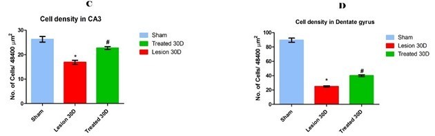
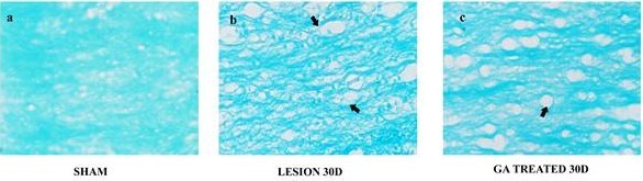
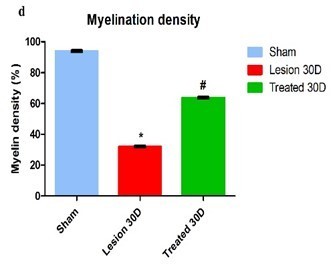
Brain sections at the level of dorsal hippocampus (Bregma -3.00 to -3.50) were stained with Luxol fast blue. Corpus callosum at this level was photomicrographed at 400x magnification. These images were then analysed offline using, Image J software (NIH, USA). In brief, the images were first converted to grey scale image using the image Statistical analysis All data were expressed as mean ± standard error mean (SEM) and the statistical analysis of the results was performed by one-way analysis of variance (ANOVA) followed by Tukey’s test using Graph Pad Prism 5. p values ≤ 0.05 was considered significant. Observations The effect of treatment of GA on the Chronic cerebral hypoperfusive rats was well documented in this study through their behaviour in NOR and HB task and the histological assessments. Novel object recognition Test On day 30 after BCCAo the Discriminative index of lesion group (0.1917±0.0305, Mean ± SEM) was significantly lower than sham group. On the other hand of treated group (0.5047±0.0268) was found to be significantly higher than lesion group (Fig 1A). Neophilia is a complex behaviour to understand, however this pathology seems to be complex because it believed that many cognitive substrates play major roles and thus when the rats experience anhedonia, they do not venture in NOR test, it is a must to mention that GA helps in the improvement of the condition and hence the rats exhibit neophilia. Hole board test The Exploratory drive was measured in terms of head dips during 5 minutes period on day 30 after BCCAo was significantly lower in lesion group (25.80±1.420) when compared to the sham group rats and the treated group (36.10±1.933) made significant increase in head dips than lesion group rats (Fig. 1B). This makes it clear that the exploratory drive follows a gradual diminishing trend, however the treatment seems to rescue. Histomorphological investigation of grey and white matter. Grey matter degeneration and consequent salvage by GA Chronic cerebral hypoperfusion (CCH) is a very peculiar and rather different form of cerebrovascular ailment where the distortion in cytoarchitecture doesn’t always follow a similar trend as that of the focal cerebral ischemia. Cresyl fast violet being the vital stain used in neuro-histology for demonstrating viable neurons was employed to determine the status of the neurons in lesion and treatment. Histological investigations of sham rat brains were made and it revealed pristine looking neurons with centrally placed pale round profiled nucleus and dark nucleolus , thin rim of cytoplasm stained bluish violet, prominent nissl substance which was stained in a similar fashion, indicating nuclear health and robustness and in no case there was any hint of chromatolyis as well. The aforementioned features were regarded as inclusion criteria for selection of viability in lesion and treated tissues for quantifying spared neurons (Fig. 3 and 4). The count of normal pyramidal neurons within the 48400µm2 of defined CA1 region of bilateral hippocampus in the sham group rats was 59.39±1.263 (Mean ± SE). The cell count in the lesion group (24.89±0.8778) was significantly reduced when compared with sham group rats and the treated group rats (40.06±1.230) show a significant increase in number than the lesion group (Fig.3A). CA2 region of bilateral hippocampus in the sham group rats was 41.72±1.631, the cell count in the lesion group (18.78±0.9273) was significantly reduced when compared with sham group rat and in the treated group (30.39±1.401) and CA2 pyramidal neurons of treated group rats were significantly higher than lesion group (Fig.3B). CA3 region of bilateral hippocampus in the sham group rats was (26.28±1.123), the cell count in the lesion group (16.89±0.8161) was significantly reduced when compared with sham group rats. However, the treated group (22.72±0.604) was significantly higher than lesion group (Fig.3C). DG region of hippocampus in the sham group rats was 89.44±2.916. The DG cell count in the lesion group (36.06±2.0070) was significantly reduced when compared with sham group rats but in the treated group (58.78±2.337) it was found to be significantly higher in number than that of the lesion group (Fig.3D). White matter derangement and concurrent restoration by GA Myelin staining reveals aqua blue coloured thick intense staining with no unstained spaces and vacuolation in sham rat brain tissue. However, in lesion it was evident that there was pericellular spacing, rarefaction of myelin bundles and was found to be reduced staining intensity in the tissue, at times looked oedematous as well. Quantification of fibre density using the Renyi entropy protocol through ImageJ was performed to substantiate the potentials of white matter salvaging activity of GA. (Fig. 4). Myelin density of the sham group rat was 93.97±0.5729, lesion group (32.07±0.5093) was severely affected than sham group; the treated group (63.72±0.5304) rats showed significant restoration in fibre density in corpus callosum, when compared with lesion group (Fig. 4 a-c). Effects of GA on chronic cerebral hypoperfusion induced neophilia and other higher cognitive deficits in rats. (A) Discriminative indices of rats after drug administration post lesioning. (B) No of head dips made during the hole board trial. Data represents Mean ± SEM of various groups which are analysed by ANOVA and compared through Turkey’s test where n=6, *p < 0.05 vs sham ; # p < 0.05 vs lesion. Effects of GA on chronic cerebral hypoperfusion induced neuropathological changes via CFV; 40x magnification of CA1 after 30 days follow up period. (2a-c) depicts 4x images of hippocampus representing the three groups and the various subfields viz. CA1, CA2, CA3, (CA- Cornu Ammonis) DG (DG-Dentate gyrus). (2d-f) 40x images illustrating viable cells (black pointed arrow) and lesioned pyknotic and shrivelled cells (black bold arrow) treated tissue exhibited coexistence of both populations. Fig.3. Effects of GA on chronic cerebral hypoperfusion induced neuropathological changes via histomorphometry quantification. A to D histograms depicting the mean no: of viable cells from the various subfields of hippocampus in the previously mentioned CFV stained sections Data represents Mean ± SEM of various groups which are analysed by ANOVA and compared through Turkey’s test where n=6, *p < 0.05 vs sham ; # p < 0.05 vs lesion. Fig. 4 Histological appearance of Corpus callosum (myelin density) in animals of experimental groups Effects of GA on chronic cerebral hypoperfusion induced myelin derangements like vacuolation, rarefaction 3(a-c) black bold arrows show regions of demyelinated, vacuolated and rarefied white matter, which is maximum in lesion panel and d) The histogram also substantiates the same through image-J quantification. Data represents Mean ± SEM of various groups which are analysed by ANOVA and compared through Turkey’s test where n=6, *p < 0.05 vs sham; # p < 0.05 vs lesion. Discussion Permanent bilateral common carotid artery occlusion as a model for chronic cerebral hypoperfusion and vascular dementia affects the brain severely with its chronic nature of insult (Wakita et al., 2002). Cerebral blood flow (CBF) pattern is quite different from that of the transient focal ischemic events, the CBF undergoes a dynamic change, initially, it drastically falls down (ischemic phase) and then the vertebro-basilar tree takes over the job of recuperating the CBF (oligaemic phase) this lasts for 8 weeks (Farkas et al., 2007). In our study Glycyrrhizic acid administration has been mainly a protective and preventive type of module for grey and white matter derangement in the 30 day study period. GA was administered through the entire period which results in alleviation of the pathology of chronic cerebral hypoperfusion. Hence, the effect of Glycyrrhizic acid has been primarily to protect the cells from oligaemic burden and shows promising inclination. Histologically it has been inferred from our work that the model has a global diffuse effect which was spanning to 30 days. The analysis of lesion group has revealed a range of deleterious changes in various areas includes striatum, hippocampal CA1, CA2, CA3 and DG. CA1 region of hippocampus is known to be the most vulnerable subgroup of cells (Niizuma et al., 2008) and it serves as an important cognitive substrate in learning and memory (Zhao et al., 2014). On the other hand, lesioning of CA2 cells causes temporal order processing deficit in Novel object recognition like paradigms. To put it simply, rats bearing CA2 lesion wouldn’t be able distinguish the novel from familiar object and they cannot process. when this novel object was introduced in the maze, this makes them explore both the objects with impaired level of curiosity, thus scoring low in discriminative index (Caruana et al., 2012). CA3 cells are specialized in place and object, place and odour association in the learning process, lesioning the CA3 greatly impairs the associative component in learning. Finally, DG as we all know is the main input for the trisynaptic network (CA3 – CA2- CA1) in hippocampus (Ji et al., 2008). Cell density in hippocampus in treated group clearly hints towards an anti-oxidant property of the drug being involved in preventing the cells from damage. GA has been shown to possess inhibiting effect on 11-β hydroxysteroid dehydrogenase (11 β HSD) enzyme activity thus exerting an anti-inflammatory effect (Ploeger 2001). This implies that it has the property of reducing inflammatory response, countering one important cascade of events that lead to oligaemic neuronal death. GA is reported to possess strong antioxidant and free radical scavenging activity (Ploeger 2001; Visavadiya et al., 2009). White matter rarefaction was also observed to be significant in lesion rats. In the lesion groups, it was observed that the corpus callosum was left with only half as much as the original degree of fibre density (Fig 4). Chronic cerebral hypoperfusion leads to constant production of ROS and this in turn results in lipid peroxidation (Farkas et al., 2007). This could be an explanation to the severe white matter rarefaction that has been observed (Shibata et al., 2004). However, GA appeared to have a significant ameliorating effect on white matter damage induced by oxidative stress (Fig 4) which was seen in our study. Loss of pyramidal neurons in the hippocampus and the behavioural changes have also been in congruence with the cell loss, which was documented with Novel Object Task (Sarti et al., 2002). The vast amount of cell death observed could be the result of a variety of cell death cascades, such as those brought about by inflammation and apoptosis, in response to the chronic ischemic insult (Bennett et al., 1998; Harukuni and Bhardwaj 2006). This change in discriminative index should be analysed from a striatal perspective because it plays a major role in executive function. The reduction in exploratory drive of the rats following lesion was recuperated after treatment with GA (Sarti et al., 2002). Treated rats showed significantly higher levels of neophilia, as compared to the lesion group. The improvement in cognitive functions after treatment with GA could be a result of restored function of NMDA receptors which are involved in excitotoxity induced by CCH. Conclusion In this study the effect of Glycyrrhizic acid on the rat model of chronic cerebral hypoperfusion was quite perceivable to be a fitting arsenal against the insult posed by chronic cerebral hypoperfusion. It is seen to improve the learning and memory function and also made consistent improvement in non-spatial memory domains and higher cognitive functions. Also reduced the count of neural death in various sub regions of hippocampus claiming a definitive role in mitigating chronic cerebral hypoperfusion mediated neurodegeneration and cognitive decline. References Alladi S, Kaul S, Meena AK, Somayajula S, Umadevi M, Reddy JM (2006) Pattern of vascular dementia in India: study of clinical features, imaging, and vascular mechanisms from a hospital dementia registry.J Stroke Cerebrovasc Dis,, 15: 49-56. Aliev G, Smith MA, Obrenovich ME, Jack C, Perry G (2003) Role of vascular hypoperfusion-induced oxidative stress and mitochondria failure in the pathogenesis of Alzheimer disease.Neurotox Res,5: 491-504. Battistin L, Cagnin A (2010) Vascular cognitive disorder. A biological and clinical overview.Neurochem Res,35: 1933-1938. Bennett SA, Tenniswood M, Chen JH, Davidson CM, Keyes MT, Fortin T, Pappas A (1998) Chronic cerebral hypoperfusion elicits neuronal apoptosis and behavioral impairment.Neuro Rep,9: 161–166. Bevins RA, Besheer J (2006) Object recognition in rats and mice: a one-trial non-matching-to-sample learning task to study recognition memory.Nat Protoc,1: 1306-1311. Blumenthal M, Goldberg A, Brinckmann J (2000) Herbal medicine: expanded Commission E monographs. Austin, TX: Integrative Medicine Communications. 237. Caruana DA, Alexander GM, Dudek SM (2012) New insights into the regulation of synaptic plasticity from an unexpected place: hippocampal area CA2.Learning & memory,19: 391-400. Dhingra D, Sharma A (2006) Antidepressant-like activity of Glycyrrhizaglabra L. in mouse models of immobility tests.Prog Neuropsychopharmacol Biol Psychiatry,30: 449–454. Farkas E, Luiten PG, Bari F (2007) Permanent, bilateral common carotid artery occlusion in the rat: a model for chronic cerebral hypoperfusion-related neurodegenerative diseases. Brain Res Rev,54:162-180. File SE, Wardill AG (1975) Validity of head-dipping as a measure of exploration in a modified hole-board. Psychopharmacol,44: 53-59. Genovese TM (2008) Glycyrrhizin reduces secondary inflammatory process after spinal cord compression injury in mice. Shock31: 367–375. Harukuni I, Bhardwaj A (2006) Mechanisms of brain injury after global cerebral ischemia.Neurol Clin,24: 1-21. Iadecola C (2013) The pathobiology of vascular dementia.Neuron,80: 844-866. Ji J, Maren S (2008) Differential roles for hippocampal areas CA1 and CA3 in the contextual encoding and retrieval of extinguished fear.Learning & Memory,15: 244-251. Kim H (2005) Neuroprotective herbs for stroke therapy in traditional eastern Medicine.Neurol Res,27: 287-301. Kim SW, Jin Y, Shin JH, Kim ID, Lee HK, Park S, Lee JK (2012) Glycyrrhizic acid affords robust neuroprotection in the post ischemic brain via anti-inflammatory effect by inhibiting HMGB1 phosphorylation and secretion.Neurobiol Dis,46: 147-156. Kim SW, Lim CM, Lee HK, Lee JK (2011) The use of Stronger Neo-Minophagen C, a glycyrrhizin-containing preparation, in robust neuroprotection in the post ischemic brain.Anat Cell Biol,44: 304-313. Ramesh Kumar R, Kathiravan K, Muthusamy R (2012) Bacopa monniera a Potent Neuroprotector against Transient Global Cerebral Ischemia Induced Hippocampal Damage and Memory Function.Int J Anat Sci,3: 26-32. Niizuma K, Endo H, Nito C, Myer DJ, Kim GS, Chan PH (2008) The PIDDosome mediates delayed death of hippocampal CA1 neurons after transient global cerebral ischemia in rats.PNAS,105: 16368-16373. Ploeger B, Mensinga T, Sips A, Seinen W, Meulenbelt J, DeJongh J (2001) The pharmaco-kinetics of glycyrrhizic acid evaluated by physiologically based pharmacokinetic modeling.Drug Metab Rev,33: 125-147. Sarti C, Pantoni L, Bartolini L, Inzitari D (2002) Persistent impairment of gait performances and working memory after bilateral common carotid artery occlusion in the adult wistar rat.Behav. Brain Res,136: 13–20. Sharifzadeh M (2008) A time course analysis of systemic administration of aqueous licorice extract on spatial memory retention in rats.Planta Med, 74: 485–90. SShibata M, Ohtani R, Ihara M, Tomimoto H (2004) White matter lesions and glial activation in a novel mouse model of chronic cerebral hypoperfusion.Stroke, 35: 2598-2603. Visavadiya NP, Narasimhacharya AV, (2006) Hypocholesterolaemic and antioxidant effects of Glycyrrhizaglabra (Linn) in rats.Mol Nutr Food Res,50:1080–1086. Visavadiya NP, Soni B, Dalwadi N (2009) Evaluation of antioxidant and anti-atherogenic properties of Glycyrrhiza glabra root using in vitro models. Int J Food Sci Nutr, 60: 135-149. Wakita H, Tomimoto H, Akiguchi I, Matsuo A, Lin JX, Ihara M, McGeer PL (2002) Axonal damage and demyelination in the white matter after chronic cerebral hypo perfusion in the rat. Brain Res,924: 63-67.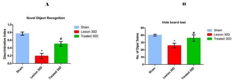
Fig. 1 Novelty seeking behaviour and Non spatial memory assessment using Novel object recognition test (A) and Exploratory behaviour assessment using Hole board test (B)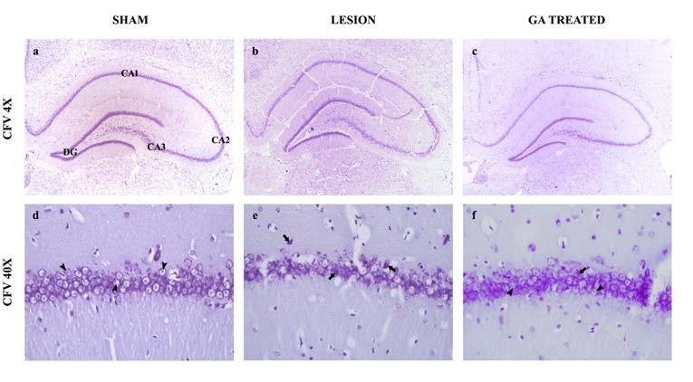
Fig. 2 Histological appearance of hippocampus in animals of experimental groups