International Journal of Anatomical Sciences 2011, 2(2):31-33
Case Report
Incomplete Horizontal Fissure of Right Lung – A Case Report
Kirubhanand C, Tamilselvi P, Sridhar Skylab R, Sankar V.
Department of Anatomy, Dr.A.L.M.Postgraduate Institute of Basic Medical Sciences, University of Madras, Taramani Campus, Chennai 600 113, Tamilnadu, India
Abstract: The right lung has two fissures, an oblique and a horizontal, dividing it into three lobes namely the upper, middle and lower. The anomaly of the fissure pattern has been described by many radiologist, whereas, there were only fewer studies on gross anatomical specimens. The present case describes a peculiar incomplete horizontal fissure which started from the oblique fissure but did not traversed backwards towards the medial surface of the lung. Anatomical knowledge of anomalies of fissures and lobes of lungs is important for surgeons performing lobectomies, radiologists interpreting X-ray and CT scans and also of academic interest to all medical personnel.
Key words: anatomical variation, abnormal lung fissure
The right lung normally has three lobes namely the upper, middle and lower formed by two fissures; an oblique and a horizontal one (Standring, 2005). The oblique fissure runs downwards, thereby meeting the inferior border of the lung at a distance of approximately 7.5 cm behind anterior end (Standring 2005). The horizontal fissure passes from the oblique fissure at the level of midaxillary line to the anterior border of the lung at the level of sternal end of fourth costal cartilage (Standring 2005). In the present case we report an incomplete horizontal fissure in the right lung specimen for its rarity and academic interest.
Case Report
During routine dissection training for postgraduate students, in a 55 year old female cadaver, we encountered an
Correspondence to: Sankar V, Department of Anatomy, Dr. ALM PGIBMS, University of Madras, Taramani Campus, Chennai 600113,Tamilnadu, India.Email: [email protected]
Accepted: 15-Sep-2011
anomalous right lung, which displayed incomplete horizontal fissure. The pulmonary fissures and lobes were studied and appropriate measurements were taken. The specimen was photographed.
The right lung displayed an oblique fissure, which originated at a distance of 7cm from the apex on the vertebral part of medial surface and after traversing a distance of 8 cm, it continued downwards as the conventional oblique fissure to cross the inferior border at a distance of 2 cm. Thus the oblique fissure was as per standard descriptions. Although the horizontal fissure was seen as per typical description i.e. it runs from oblique fissure 10 cm from the anterior border, it did not traverse backwards towards the medial surface of the lung. Thus, instead of traversing the entire lung, the horizontal fissure seen in this specimen did not divide the lung completely into a middle lobe. As a consequence to such anomalous incomplete horizontal fissure, the right lung was found to possess a completely divided lower lobe with upper and middle lobes incompletely separated from each other. No abnormality was detected in the left lung.
Fig. 1 Photograph showing the right lung seen with an incomplete horizontal fissure
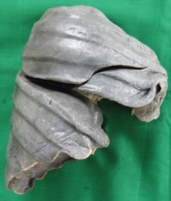
Fig. 2 Diagrammatic representation of fissure pattern of normal right lung and the anomalous one reported

Discussion
Lung develops from numerous bronchopulmonary buds which fuse completely in the later part of development except at sites of fissure formation, resulting in the formation of lobes and fissures (Frija et al., 1988). Any deviation from the normal pathway of fusion of bronchopulmonary buds results in the formation of variations involving lobes and fissures of the lungs (Sadler, 2004). The fissures are the spaces which separate individual bronchopul- monary buds or segments and they get obliterated except along the two planes which later manifests as horizontal or oblique fissure Non-obliteration of these spaces gives rise to accessory issures of the lung (Meenakshi, 2004).
An incomplete fissure may be of varying depth occurring between bronco- pulmonary segments and is also a cause for post operative air leakage (Walker, 1997). Often accessory fissures act as barriers to infection spread, creating a sharply marinated pneumonia which can wrongly be interpreted as atelectasis or consolidation (Godwin and Tarver, 1984). The knowledge of anatomy of fissures of lung may help clarifying initially confusing radiographic findings like extension of fluid into an incomplete major fissure or spread of various diseases through different pathways (Dandy, 1978) and explain radiological appearances of interlobar fluid (Raasch,1982). Accurate recognition of lung anomalies in different populations will improve the understanding of lesions like pneumonia, pleural effusion, and collateral air drift along with disease spreading through the lung.
Aldur et al., (1997) concluded that a surgeon must always remember the anatomical variations of the location of the lungs especially in lobectomies and in segmental resection. Hayashi et al., (2001) concluded that anatomy of normal variants of the major fissures is essential for recognizing their variable imaging appearances and related abnormalities.
The presence of fissures in the normal lungs enhances uniform expansion
Kirubhanand et al., – Incomplete horizontal fissue of right lung and hence facilitates more air intake. Accessory and incomplete fissures of varying depth can be seen in unusual Standring S (2005) Gray’s Anatomy. 39th New York: Churchill Livingstone,.
Edition.locations of the lung, delimiting abnormal lobes which corresponding to the normal bronchopulmonary segments especially in infants. From a radiological point of view, an accessory or anomalous fissure is important as it can be mistaken for a lung lesion or an atypical appearance of pleural effusion.
Considering the clinical and surgical importance of such variations, from anatomical point of view, one can opine that prior anatomical knowledge and high index of suspicion for probable variations in the fissures, lobes and bronchopulmonary segments in the lung may be important for clinicians, surgeons and radiologists.
References
Aldur MM, Denk CC, Celik HH (1997) An accessory fissure in the lower lobe of the right lung. Morphologie, 81: 5-7.
Aziz A, Ashizawa K, Nagaoki K (2004) High resolution CT anatomy of the pulmonary fissures. J Thorac Imaging, 19: 186-191.
Dandy WE (1978) Incomplete pulmonary interlobar fissure sign. Radiology, 128: 21-25.
Godwin JD, Tarver RD (1984) Accessory Fissures of the Lung. AJR, 144: 39-47.
Frija J, Naazib J, David M, Hacein-Bey L, Yana C, Laval-Jeantet M (1988) Incomplete and accessory pulmonary fissures studied by high resolution x-ray computed tomography. J Radiol, 69: 163-170.
Hayashi K, Aziz A, Ashizawa K, Hayashi H, Nagaoki K, Otsuji H (2001) Radiographic and CT appearances of the major fissures. Radiographics, 21: 861-874.
Meenakshi S, Manjunath KY, Balasubramanyam V (2004) Morphological variations of the Lung Fissures and lobes. The Indian J Chest Dis & Allied Sci, 46: 179-178.
Raasch BN, Carsky EW, Lane EJ, O’Callaghan JP, Heitzman ER (1982) Radiographic anatomy of the interlobar fissures: a study of 100 specimens. Am J Roentgenol, 138: 1043-1049.
Saddler TW (2004) Langman’s Medical Embryology.9th Edition. Baltimore: Lippincott Williams & Wilkins.
Walker WS, Craig SR (1997) A proposed anatomical classification of the pulmonary fissures. J R Coll Surg Edinburg, 42: 233-234.
Kirubhanand et al., – Incomplete horizontal fissue of right lung